Iowa Eye Association News
Oct. 2004. Series 2, no. 39.
Iowa Eye Association News Oct. 2004. Series 2, no. 39. |
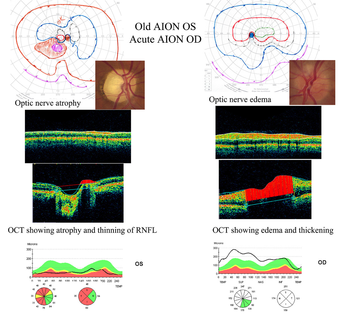
Figure 1. This figure shows a composite of visual fields (Goldmann), optic nerve photos, and optical coherence tomography (OCT) of the optic nerve head (sagital sections from nerve head scan) and retinal nerve fiber layer thickness (circular RNFL scan) in a patient with old anterior ischemic optic neuropathy (AION) OS and acute AION OD.
Note that in the left eye there is a pale nerve, inferior altitudinal visual field loss that is greater than the superior loss, and acquired cupping of the nerve head on OCT and thinning of the retinal nerve fiber layer both in the inferior and superior bundles (worse in the superior bundle, corresponding to the inferior field loss). The red pie-shaped segments of the retinal nerve fiber layer represent significant thinning at the 1% probability level compared to age-matched normals (yellow is the 5% significance level).
In the right eye with acute AION, there is optic disc edema and abnormal thickening of the retinal nerve fiber layer, which will eventually thin and appear similar to the left eye over time.
(use your browser's scroll bar to view image)
|
(requires Adobe Acrobat Reader) |
|||
| "Together,
we have a vision for the future." |
Copyright © University
of Iowa, 2004 |
||