Case Studies in Ophthalmology
Cases for the Ophthalmology Clerkship
Note to Medical Students on the Ophthalmology Rotation at the University of Iowa:
Print the question and answer sheet (Ctrl-Print or Command-Print) and enter your answers on it. You may prefer to type your answers on this page and then print it. If you do this, be careful that your full answer shows in the printout. Experience with Internet Explorer has been less than optimal.
Return the printed copy with your answers to Michelle Snyder or Dr. Kemp via campus mail (11290 PFP) or scan as a pdf and email to Michelle at michelle-r-snyder@uiowa.edu or Dr. Kemp at pavlina-kemp@uiowa.edu.
Case #18 Optic Atrophy
History
Patient is a 70-year-old man who is in good health and presents with a history of sudden visual loss in the left eye. When carefully questioned, he says that he actually discovered poor vision in the left eye when he was poked in the right eye by his baby grandson and rubbed the right eye.
Photo #1
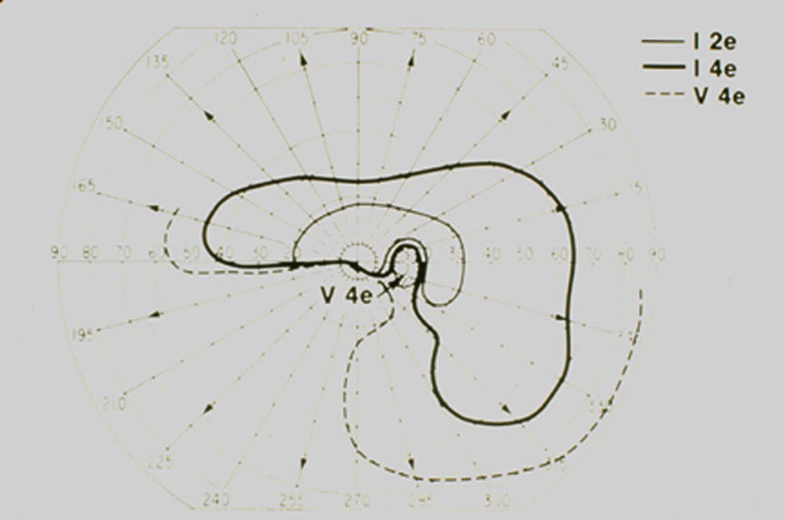
Photo #2
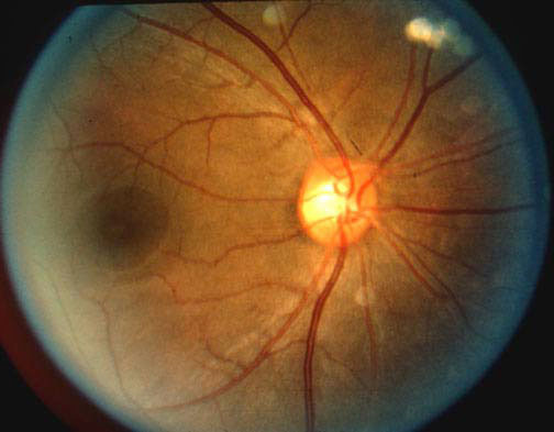
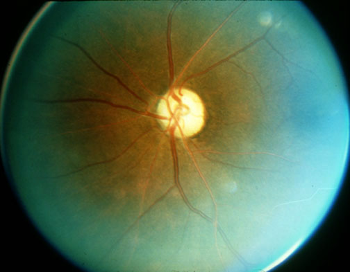
Photo #3
Enter Full Name (required):
Your email address (required):
Question #1
Visual acuity was 20/200 OD and there was a 1.2 log unit relative afferent pupillary defect in the left eye.
Question #2
What form of optic nerve damage must be ruled out?
Question #3
What differential diagnosis is realistic at this age?
Question #4
Why would you get a sedimentation rate?
Question #5
What laboratory and radiographic studies would you want to obtain?
Question #6
What is the reason for the relative afferent pupillary defect?
Question #7
What are the pathogenetic mechanisms of visual loss with optic nerve compression?
Question #8
What are the commonest causes of compression of the optic nerve?
Question #9
What is the characteristic first visual field defect with optic nerve compression? How does it progress?
Question #10
If the patient had a central scotoma in the right eye and a superior temporal defect in the left, where would the lesion be located? What is this combination of field defects called?
References
Hayreh SS: Anterior ischemic optic neuropathy. Arch Neurol 1981; 38: 675-678.
Beck RW, et al.: Optic disc structure in anterior ischemic optic neuropathy. Ophthalmology 1984; 91:1334-1337.
Hamilton CR, et al.: Giant cell arteritis: including temporal arteritis and polymyalgia rheumatica. Meidicine 1971; 50:1-30.
McDonal WI: The symptomatology of tumors of the anterior visual pathways. Canad J Neurol Sci 1982; 9:381-390.
Spencer WH: Primary neoplasms of the optic nerve and its sheaths: clinical features and current concepts of the pathogenetic mechanism. Trans Am Ophthalmol Soc 1972; 70:490-528.