Case Studies in Ophthalmology
Cases for the Ophthalmology Clerkship
Note to Medical Students on the Ophthalmology Rotation at the University of Iowa:
Print the question and answer sheet (Ctrl-Print or Command-Print) and enter your answers on it. You may prefer to type your answers on this page and then print it. If you do this, be careful that your full answer shows in the printout. Experience with Internet Explorer has been less than optimal.
Return the printed copy with your answers to Michelle Snyder or Dr. Kemp via campus mail (11290 PFP) or scan as a pdf and email to Michelle at michelle-r-snyder@uiowa.edu or Dr. Kemp at pavlina-kemp@uiowa.edu.
Case #4 Nonproliferative Diabetic Retinopathy
History
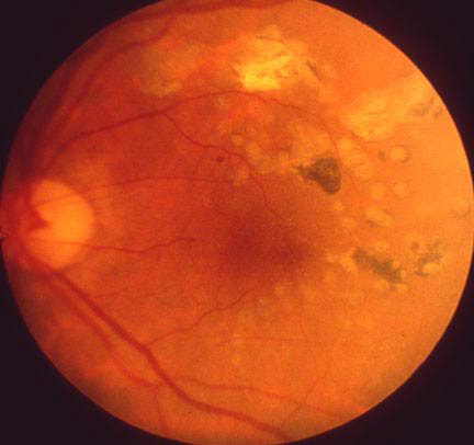
58-year-old maturity onset diabetic presents with a 6 month history of gradually progressive visual loss in the left eye. The first photograph is a fundus photo taken on the day of presentation. Two years later the ocular fundus was photographed again.
Photo #1
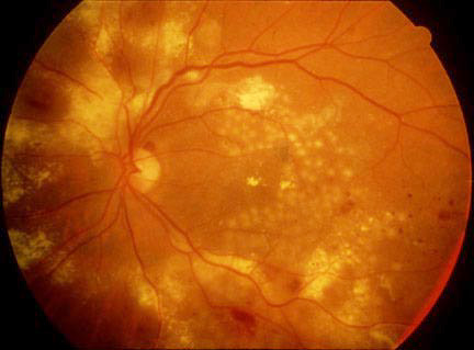
Photo #2 (Two Years Later)
Enter Full Name (required):
Your email address (required):
Question #1
What are the findings in the left fundus and what is your diagnosis?
Question #2
What is the yellow substance within the retina and what caused it?
Question #3
What questions would you ask the patient? What other things would you look for on the physical examination or laboratory studies?
Question #4
What is the appropriate treatment for the patient? Is any medical treatment effective?
Question #5
What is the long-term prognosis for this patient?
References
Fine SL, Patz A: Sights and Sounds in Ophthalmology. Vol 4. Diabetic Retinopahty. A slide-tape presentation of the Retinal Vascular Center, Wilmer Institute, Mosby.
Early Treatment Diabetic Retinopathy Study Research Group: Early treatment diabetic retinopathy study: Report I: Photocoagulation for Diabetic Macula Edema. Arch Ophthalmol 1885; 103:796-806.