Case Studies in Ophthalmology
Cases for the Ophthalmology Clerkship
Note to Medical Students on the Ophthalmology Rotation at the University of Iowa:
Print the question and answer sheet (Ctrl-Print or Command-Print) and enter your answers on it. You may prefer to type your answers on this page and then print it. If you do this, be careful that your full answer shows in the printout. Experience with Internet Explorer has been less than optimal.
Return the printed copy with your answers to Michelle Snyder or Dr. Kemp via campus mail (11290 PFP) or scan as a pdf and email to Michelle at michelle-r-snyder@uiowa.edu or Dr. Kemp at pavlina-kemp@uiowa.edu.
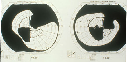
Case #49 Field Defects
History
The patient is a 45 year old patient with visual field loss.
Enter Full Name (required):
Your email address (required):
Question #1
How could you account for her complaint that her vision is getting worse?
Question #2
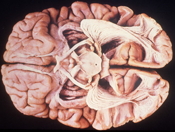
Where is/are the lesions likely to be in order to produce the field loss? (please describe each below, brain image is included for your information and is (obviously) not that of the patient.)
a) at the nerve head(s) and at the chiasm
b) at both nerve head(s) and in the right occipital lobe
c) in both occipital lobes
d) at both nerve heads
e) in the optic radiations
References
Glubovovicz and Williams: The Human Brain, A Photographic Guide. Harper & Row, Hagerstown, MD 1980.
Walsh FB, Hoyt WF: Clinical Neuro-Ophthalmology 3rd Ed. Williams & Wilkins, Baltimore, MD pp 43-96; 1969.