This site uses tracking information. Visit our privacy policy. Click to agree to this policy and not see this again.

Ophthalmology and Visual Sciences

Anterior Ischemic Optic Neuropathy:
Part I: An Introduction for Patients

Anterior Ischemic Optic Neuropathy:
Part I: An Introduction for Patients

Category(ies): Neuro-ophthalmology
Contributors: Sohan Singh Hayreh, MD, MS, PhD, DSc, FRCS, FRCOphth
Patricia A. Podhajsky, BSN

Ocular Vascular Clinic
Department of Ophthalmology & Visual Sciences
University of Iowa Carver College of Medicine
Iowa City, Iowa

This is part one in a pair of articles about AION.

PLEASE NOTE

The first part of this article is intended primarily for patients and lay people, and the second part (on a separate web page) is intended for ophthalmologists. The article is a summary of material published in peer-reviewed ophthalmic journals. For more detailed information, please refer to the papers in the bibliography and the various articles cited in those papers.

Dr. Hayreh does not give an opinion without personally examining a patient; he feels that to do so is unethical and also potentially dangerous.

Originally published 1995 and revised, expanded and modified in April 2009, reviewed November 2010, February 2013, August 2016.

This article is intended to give basic information about ischemic optic neuropathy, which is sometimes called "stroke of the optic nerve" in lay persons' term. Ischemic optic neuropathy is one of the most common causes of loss of vision in persons past middle age. However, it does occur less commonly, in younger persons. Knowledge about this disease will enable patients and their doctors to work together to understand the disease. Early diagnosis, recognition of various risk factors, and carrying out the treatment and follow-up visits as recommended, are the most important aspects of care for persons with ischemic optic neuropathy. The following description of ischemic optic neuropathy is based on our basic, experimental and clinical studies published in the peer reviewed journals. [1-77]


Background Information

The Eye

The eye is often compared to a camera. Light comes in through the cornea, pupil, and lens at the front of the eye just as the lens of the camera lets in light to the film. This light is then focused on the inside wall of the eye called the retina (as on the film in a camera). This picture is then sent to the brain along the optic nerve, which connects the eye to the brain. We see properly when all of these parts of the eye and brain are working together

The Optic Nerve

The optic nerve is unique because it is the only nerve in the body that a physician is able to see directly (without surgery) as it begins at the back of eye. The optic nerve is made up of a million tiny, delicate nerve fibers that are like wires. Many blood vessels nourish the optic nerve with blood rich in oxygen and nutrition. Vision actually takes place in the brain when the messages from the eye travel to the brain along the optic nerve; however, the nerve has to be healthy to transmit these messages. The whole situation may be compared to a video camera (i.e. the eye) taking the picture and transmitting along the cable (the optic nerve) to the television (the brain); if the cable is damaged for any reason, although the video camera and the television may be perfectly normal, a distorted picture or no picture is seen on the television.

While explaining this disease, a doctor may say that there has been a "stroke at the back of the eye", "blocked blood vessel", or "broken blood vessel at the back of the eye". A "stroke" is an interruption of the blood supply to the brain or a nerve in the body. When a person has a major "stroke" in the brain, the blood supply to a part of the brain that controls speech or movement of the arms and legs is often affected. With a "stroke at the back of the eye" a loss of vision develops because the retina or the optic nerve may have its circulation interrupted. How much vision is lost depends on how much the retina or the optic nerve is affected. In this website we are discussing only the stroke of the optic nerve and NOT that of the retina.

Ischemia

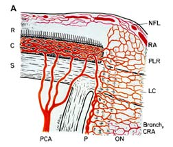
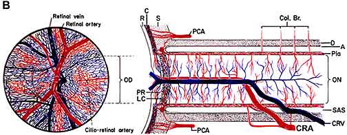
Ischemia of the optic nerve results in ischemic optic neuropathy [17, 24, 36, 49, 57, 74]. Ischemia is a decrease in the blood supply to a tissue in the body which in turn decreases oxygen and nutrition. Without sufficient oxygen and nutrition, nerves cannot function properly and will eventually die. As an example, placing a tight rubber band around the wrist causes the fingers to begin to tingle and then lose the sensation of touch because the rubber band cuts off the blood supply, starving the nerves of oxygen and nutrients. If the blood supply to the optic nerve is interrupted, it produces ischemia of the optic nerve (technical name, "ischemic optic neuropathy"), and a loss of vision develops. The blood supply of the anterior (Fig. 1-A) of the optic nerve (also called the optic nerve head) is mainly by the posterior ciliary arteries, while the blood supply to the posterior part (back part of Fig. 1-B) is from several quite different sources but not the posterior ciliary arteries [4-6, 8, 9, 13, 18, 25, 30, 35, 36, 49, 52, 74].

Schematic Representation Of Blood Supply of: (A) The Optic Nerve Head
Fig. 1a: Schematic Representation Of Blood Supply of: (A) The Optic Nerve Head. A Reproduced With Permission From Hayreh 1978 [16]. B Modified From Hayreh, S.S. 1974 [8].
Abbreviations: A = arachnoid; C= choroid; CRA= central retinal artery; Col. Br.= Collateral branches; CRV= central retinal vein; D= dura; LC= lamina cribrosa; NFL= surface nerve fiber layer of the disc; OD= optic disc; ON= optic nerve; P= pia; PCA= posterior ciliary artery; PR and PLR= prelaminar region; R= retina; RA= retinal arteriole; S= sclera; SAS= subarachnoid space.
Schematic Representation Of Blood Supply of: (A) The Optic Nerve Head
Fig. 1b: Schematic Representation Of Blood Supply of: ((B) The Optic Nerve. A Reproduced With Permission From Hayreh 1978 [16]. B Modified From Hayreh, S.S. 1974 [8].
Abbreviations: A = arachnoid; C= choroid; CRA= central retinal artery; Col. Br.= Collateral branches; CRV= central retinal vein; D= dura; LC= lamina cribrosa; NFL= surface nerve fiber layer of the disc; OD= optic disc; ON= optic nerve; P= pia; PCA= posterior ciliary artery; PR and PLR= prelaminar region; R= retina; RA= retinal arteriole; S= sclera; SAS= subarachnoid space.

Ischemia

Ischemic optic neuropathy is of two types [17, 36, 74], depending upon the part of the optic nerve involved:

Anterior ischemic optic neuropathy (commonly abbreviated to "AION"): AION is due to acute ischemia of the front (anterior) part of the optic nerve (also called optic nerve head), which is supplied mainly by the posterior ciliary arteries.

Posterior ischemic optic neuropathy (commonly abbreviated to "PION"): PION is a much less common type [24, 60]. It is due to acute ischemia of the back (posterior) part of the optic nerve, located some distance behind the eyeball; this part of the optic nerve is NOT supplied by the posterior ciliary arteries. This was first reported by me in 1981 [24]. A detailed current account of PION and its management is available in a recent publication based on my research [60].


Clinical Classification Of AION

AION is of two types [29, 36, 49, 74], depending on what causes it:

Arteritic AION: This is the most serious type and is due to a disease called giant cell arteritis or temporal arteritis.

Non-arteritic AION:: This is the usual, most common type, with many different causes but not associated with giant cell arteritis.

IT IS EXTREMELY IMPORTANT TO KNOW THAT THE CORRECT TREATMENT FOR AION DEPENDS ON WHETHER IT IS THE ARTERITIC OR THE NON-ARTERITIC TYPE.

How AION is Diagnosed

To make a correct diagnosis of AION and its type, the doctor needs to find out all the required information. He/she might ask the following questions, because painless, loss of vision due to AION may be the first symptom of, or a complication of other diseases.

QUESTIONS FOR PATIENTS WITH AION

Non-Arteritic AION

      Was the loss of vision sudden or gradual?
      If the visual loss came on suddenly, at what time of the day did the patient discover it?
      Was there temporary blurring or loss of vision before the visual loss?
      Has the visual loss been stable since first discovery, or improved or worsened?
      Is the patient diabetic?
      Has the patient had any heart trouble, stroke, high blood pressure, low blood pressure or shock, recent cardiovascular surgery or cataract surgery, or excessive bleeding?
      Does the patient or anyone in the patient's family have glaucoma?
      Does the patient smoke?
      Is the patient on high blood pressure medication? If so, which drug(s), how often and what time of the day?
      Women will be asked about use of birth control pills or estrogens used after menopause.

Arteritic AION

Giant cell arteritis is the prime medical emergency in ophthalmology because of its dreaded complication of visual loss in one or both eyes, which is preventable if these patients are diagnosed early and treated immediately and aggressively with systemic corticosteroids. This is a disease of persons aged 50 years or older - more common among women than men [40]. About 80% of patients with AION due to giant cell arteritis will have felt generally unwell for some time [40, 42]. However, 20% have no such general symptoms and are perfectly healthy [42]. Some patients may have a feeling of "euphoria," or well-being, so that they are quite unconcerned about their loss of vision and even insist they can see quite well when it is clear that they can't. Patients feeling like this may want to refuse treatment aimed at stopping the progress of giant cell arteritis and blindness, so it is important to have the cooperation and understanding of all family members. To help determine if giant cell arteritis is present, a physician will want to know about the following problems:

  • Pain while chewing food
  • Pain in the temples and/or neck
  • Scalp tenderness
  • Loss of appetite
  • Loss of weight
  • Headaches
  • Unusual fatigue or sleepiness
  • Muscular aches and pains
  • Regarding the loss of vision, the doctor might ask the same questions as those listed above in non-arteritic AION.

Tests and Procedures that may be done in patients with AION

Visual acuity testing: The ophthalmologist needs to know at every visit just how much the patient can see, so that changes in the amount of affected vision can be determined. In addition to reading the eye charts for distance and near visual acuity, patients will often have visual field testing.

Visual field testing: A visual field test measures both central (straight ahead) and peripheral (side) vision. A defect in the visual field test shows which optic nerve fibers are affected by the disease An eye with normal 20/20 (or 6/6) visual acuity can have fairly marked visual field loss in the periphery, so a normal visual acuity does not guarantee that there is no visual loss.

Relative afferent pupillary defect: The pupil, located in the center of the iris, is the part of the eye which gets larger and smaller according to the amount of light. The optic nerve plays an important role in this reaction to light, so if the pupil is constricting to light less than normal it shows that there is damage to the optic nerve or the retina. The pupils are tested by shining a light into each eye, causing the pupils to constrict. As the light is moved from one eye to the other, the pupils should constrict to about the same size. If they do not, a relative afferent pupillary defect is present in the involved eye.

Tonometry (glaucoma testing): The pressure in the eye (intraocular pressure) is measured with an instrument called a tonometer. This simple, painless procedure is essential to obtain full information about the circulation in the eye. If there is any doubt about whether the pressure is high the patient may need to be tested several times over a whole day, because intraocular pressure can vary at different times of the day. A person may not know he/she has high pressure in the eye until after visual loss has occurred.

Fundus photograph and fluorescein fundus angiography: Photographs of the optic disc and the back of the eye may be taken on several occasions during the course of the disease to find out the amount of swelling of the optic disc and its resolution. Fluorescein fundus angiography is a routine dye test, done to find out the state of circulation in the back of the eye and optic disc.


More information

More about fundus photography

More about fluorescein angiography

Blood tests and referrals to specialists in other areas: Blood tests which will be done immediately are the Erythrocyte Sedimentation Rate (ESR) and C-reactive protein (CRP). The results of these tests are available within a couple of hours. These two blood tests are extremely important to find out if a patient has giant cell arteritis. Both are usually abnormally high in giant cell arteritis.

Many other blood studies may be needed to find out if there is anything else wrong with the patient, and may help in finding the reason for the development of AION, such as diabetes or collagen vascular disease. Cholesterol and/or triglyceride levels will be checked because high levels lead to "hardening of the arteries." The results of these blood studies may indicate a need for follow-up by a local physician or referral to a specialist in hematology.

Blood pressure will be taken to determine if the patient has high blood pressure (hypertension). A cardiologist or cardiovascular specialist may be consulted if it seems that the cause of AION is a blood clot from somewhere else in the body, or that the heart, carotid arteries, or generalized "hardening of the arteries" may be contributing to the ischemia in the eye. Our recent studies have shown that abnormal fall of blood pressure during sleep is a serious risk factor for AION in the vast majority [12, 14, 15, 52, 60]. This can be tested by recording the blood pressure every 10 to 20 minutes over a 24 hour period, with an ambulatory blood pressure monitor

Temporal artery biopsy: When the patient's symptoms, the eye examination, elevated ESR and CRP, and fluorescein angiography make it seem likely that he/she has giant cell arteritis, a temporal artery biopsy will be done. The temporal artery lies just under the skin on the side of the forehead, or temple. The area is anesthetized and a small cut made in the skin to expose the artery. About an inch of the artery is removed for examination and the area is sutured with several stitches (which will be removed in about a week). This sample of the artery is examined under a microscope by a pathologist to determine whether there is inflammation of the artery. It may take several days before this result is known; however, treatment for giant cell arteritis may be started immediately if there is a strong suspicion of giant cell arteritis, even before the biopsy is done, because of the very high risk of blindness if adequate treatment is not given soon enough. Starting treatment before the biopsy is done does not interfere with the results.


Selected References

  1. Singh (Hayreh) S, Dass R (1960) The central artery of the retina. I. Origin and course. The British journal of ophthalmology 44: 193-212
  2. Singh (Hayreh) S, Dass R (1960) The central artery of the retina. II. A study of its distribution and anastomoses. The British journal of ophthalmology 44: 280-299
  3. Hayreh SS (1962) The Ophthalmic Artery: III. Branches. The British journal of ophthalmology 46: 212-247
  4. Hayreh SS (1963) The blood supply and vascular disorders of the optic nerve. An Inst Barraquer 4: 7-109
  5. Hayreh SS (1963) The Central Artery of the Retina. Its Role in the Blood Supply of the Optic Nerve. The British journal of ophthalmology 47: 651-663
  6. Hayreh SS (1969) Blood supply of the optic nerve head and its role in optic atrophy, glaucoma, and oedema of the optic disc. The British journal of ophthalmology 53: 721-748
  7. Hayreh SS, Baines JA (1972) Occlusion of the posterior ciliary artery. 3. Effects on the optic nerve head. The British journal of ophthalmology 56: 754-764
  8. Hayreh SS (1974) Anatomy and physiology of the optic nerve head. Transactions - American Academy of Ophthalmology and Otolaryngology 78: OP240-254
  9. Hayreh SS (1974) Anterior ischaemic optic neuropathy. I. Terminology and pathogenesis. The British journal of ophthalmology 58: 955-963
  10. Hayreh SS (1974) Anterior ischaemic optic neuropathy. II. Fundus on ophthalmoscopy and fluorescein angiography. The British journal of ophthalmology 58: 964-980
  11. Hayreh SS (1974) Anterior ischaemic optic neuropathy. III. Treatment, prophylaxis, and differential diagnosis. The British journal of ophthalmology 58: 981-989
  12. Hayreh SS (1974) Pathogenesis of cupping of the optic disc. The British journal of ophthalmology 58: 863-876
  13. Hayreh SS (1975) Anterior ischemic optic neuropathy. Springer-Verlag, Heidelberg
  14. Hayreh SS (1977) Optic disc edema in raised intracranial pressure. V. Pathogenesis. Archives of ophthalmology 95: 1553-1565
  15. Hayreh SS (1977) Optic disc edema in raised intracranial pressure. VI. Associated visual disturbances and their pathogenesis. Archives of ophthalmology 95: 1566-1579
  16. Hayreh SS (1978) Fluids in the anterior part of the optic nerve in health and disease. Survey of Ophthalmology 23: 1-25
  17. Hayreh SS (1978) Ischemic optic neuropathy. International Ophthalmology 1: 9-18
  18. Hayreh SS (1978) Structure and blood supply of the optic nerve. In: Heilmann K, Richardson KT (eds) Glaucoma: Conceptions of a Disease Pathogenesis, Diagnosis, and Therapy. Thieme, Stuttgart, pp. 78-96.
  19. Hayreh SS, Podhajsky PA (1979) Visual field defects in anterior ischemic optic neuropathy. Documenta Ophthalmologica Proceedings Series 19: 53-71
  20. Hayreh SS (1980) Anterior ischemic optic neuropathy. IV. Occurrence after cataract extraction. Archives of ophthalmology 98: 1410-1416
  21. Hayreh SS (1981) Anterior ischemic optic neuropathy. Archives of Neurology 38: 675-678
  22. Hayreh SS (1981) Anterior ischemic optic neuropathy. V. Optic disc edema an early sign. Archives of ophthalmology 99: 1030-1040
  23. Hayreh SS, Zahoruk RM (1981) Anterior ischemic optic neuropathy. VI. In juvenile diabetics. Ophthalmologica 182: 13-28
  24. Hayreh SS (1981) Posterior ischemic optic neuropathy. Ophthalmologica 182: 29-41
  25. Hayreh SS (1985) Inter-individual variation in blood supply of the optic nerve head. Its importance in various ischemic disorders of the optic nerve head, and glaucoma, low-tension glaucoma and allied disorders. Documenta ophthalmologica 59: 217-246
  26. Hayreh SS (1986) Anterior ischemic optic neuropathy. Japanese Journal of Clinical Ophthalmology 40: 581-587
  27. Hayreh SS, Servais GE, Virdi PS (1986) Fundus lesions in malignant hypertension. V. Hypertensive optic neuropathy. Ophthalmology 93: 74-87
  28. Hayreh SS (1987) Anterior ischemic optic neuropathy. VIII. Clinical features and pathogenesis of post-hemorrhagic amaurosis. Ophthalmology 94: 1488-1502
  29. Hayreh SS (1990) Anterior ischaemic optic neuropathy. Differentiation of arteritic from non-arteritic type and its management. Eye (London, England) 4: 25-41
  30. Hayreh SS (1990) In vivo choroidal circulation and its watershed zones. Eye (London, England) 4: 273-289
  31. Hayreh SS (1990) The role of optic nerve sheath fenestration in management of anterior ischemic optic neuropathy. Archives of ophthalmology 108: 1063-1065
  32. Hayreh SS (1991) Ophthalmic features of giant cell arteritis. Baillieres Clinical Rheumatology 5: 431-459
  33. Hayreh SS, Zimmerman MB, Podhajsky P, Alward WL (1994) Nocturnal arterial hypotension and its role in optic nerve head and ocular ischemic disorders. American journal of ophthalmology 117: 603-624
  34. Hayreh SS, Joos KM, Podhajsky PA, Long CR (1994) Systemic diseases associated with nonarteritic anterior ischemic optic neuropathy. American journal of ophthalmology 118: 766-780
  35. Hayreh SS (1995) The 1994 Von Sallman Lecture. The optic nerve head circulation in health and disease. Experimental eye research 61: 259-272
  36. Hayreh SS (1996) Acute ischemic disorders of the optic nerve: pathogenesis, clinical manifestations and management. Ophthalmology Clinics of North America 9: 407-442
  37. Hayreh SS (1996) Anteriore ischamische Optikusneuropathie. Stellenwert der nachtlichen arteriellen Hypotonie. [Anterior ischemic optic neuropathy: Role of nocturnal arterial hypotension]. Klinische Monatsblatter fur Augenheilkunde 208: aA12-17
  38. Hayreh SS (1996) Duke-elder lecture. Systemic arterial blood pressure and the eye. Eye (London, England) 10: 5-28
  39. Hayreh SS (1997) Anterior ischemic optic neuropathy. Clinical neuroscience 4: 251-263
  40. Hayreh SS, Podhajsky PA, Raman R, Zimmerman B (1997) Giant cell arteritis: validity and reliability of various diagnostic criteria. American journal of ophthalmology 123: 285-296
  41. Hayreh SS, Podhajsky PA, Zimmerman B (1997) Nonarteritic anterior ischemic optic neuropathy: time of onset of visual loss. American journal of ophthalmology 124: 641-647
  42. Hayreh SS, Podhajsky PA, Zimmerman B (1998) Occult giant cell arteritis: ocular manifestations. American journal of ophthalmology 125: 521-526
  43. Hayreh SS, Podhajsky PA, Zimmerman B (1998) Ocular manifestations of giant cell arteritis. American journal of ophthalmology 125: 509-520
  44. Hayreh SS, Podhajsky P, Zimmerman MB (1999) Beta-blocker eyedrops and nocturnal arterial hypotension. American journal of ophthalmology 128: 301-309
  45. Hayreh SS (1999) Retinal and optic nerve head ischemic disorders and atherosclerosis: role of serotonin. Progress in retinal and eye research 18: 191-221
  46. Hayreh SS, Podhajsky P, Zimmerman MB (1999) Role of nocturnal arterial hypotension in optic nerve head ischemic disorders. Ophthalmologica 213: 76-96
  47. Hayreh SS (1999) Role of nocturnal arterial hypotension in the development of ocular manifestations of systemic arterial hypertension. Current opinion in ophthalmology 10: 474-482
  48. Hayreh SS (2000) Does Levodopa improve visual function in NAION? Ophthalmology 107: 1434-1438
  49. Hayreh SS (2000) Ischaemic optic neuropathy. Indian journal of ophthalmology 48: 171-194
  50. Hayreh SS (2000) Steroid therapy for visual loss in patients with giant-cell arteritis. Lancet 355: 1572-1573
  51. Hayreh SS (2001) Blood flow in the optic nerve head and factors that may influence it. Progress in retinal and eye research 20: 595-624
  52. Hayreh SS (2001) The blood supply of the optic nerve head and the evaluation of it - myth and reality. Progress in retinal and eye research 20: 563-593
  53. Hayreh SS, Podhajsky PA, Zimmerman B (2001) Ipsilateral recurrence of nonarteritic anterior ischemic optic neuropathy. American journal of ophthalmology 132: 734-742
  54. Hayreh SS, Jonas JB (2001) Optic disc morphology after arteritic anterior ischemic optic neuropathy. Ophthalmology 108: 1586-1594
  55. Hayreh SS (2002) Diabetic papillopathy and nonarteritic anterior ischemic optic neuropathy. Survey of Ophthalmology 47: 600-602
  56. Hayreh SS, Zimmerman B, Kardon RH (2002) Visual improvement with corticosteroid therapy in giant cell arteritis. Report of a large study and review of literature. Acta ophthalmologica Scandinavica 80: 355-367
  57. Hayreh SS (2002) Neuropatías Ópticas Isquémicas. In: Arruga J, Sánchez B (eds) Neuropatías Ópticas: Diagnóstico Y Tratamiento LXXVIII Ponencia Oficial Soc Espãn Oftalmol. Soc Espãn Oftalmol, pp. 207-237.
  58. Hayreh SS, Zimmerman B (2003) Management of giant cell arteritis. Our 27-year clinical study: new light on old controversies. Ophthalmologica J 217: 239-259
  59. Hayreh SS, Zimmerman B (2003) Visual deterioration in giant cell arteritis patients while on high doses of corticosteroid therapy. Ophthalmology 110: 1204-1215
  60. Hayreh SS (2004) Posterior ischaemic optic neuropathy: clinical features, pathogenesis, and management. Eye (London, England) 18: 1188-1206
  61. Hayreh SS (2005) Erectile dysfunction drugs and non-arteritic anterior ischemic optic neuropathy: is there a cause and effect relationship? Journal of Neuro-Ophthalmology 25: 295-298
  62. Hayreh SS, Zimmerman B (2005) Visual field abnormalities in nonarteritic anterior ischemic optic neuropathy: their pattern and prevalence at initial examination. Archives of ophthalmology 123: 1554-1562
  63. Hayreh SS (2006) Amiodarone, erectile dysfunction drugs, and non-arteritic ischemic optic neuropathy. Journal of Neuro-Ophthalmology 26: 154-155
  64. Hayreh SS, Zimmerman MB (2007) Incipient nonarteritic anterior ischemic optic neuropathy. Ophthalmology 114: 1763-1772
  65. Hayreh SS, Jonas JB, Zimmerman MB (2007) Nonarteritic anterior ischemic optic neuropathy and tobacco smoking. Ophthalmology 114: 804-809
  66. . Hayreh SS, Zimmerman MB (2007) Optic disc edema in non-arteritic anterior ischemic optic neuropathy. Graefes Archive for Clinical and Experimental Ophthalmology 245: 1107-1121
  67. Hayreh SS, Fingert JH, Stone E, Jacobson DM (2008) Familial non-arteritic anterior ischemic optic neuropathy. Graefes Archive for Clinical and Experimental Ophthalmology 246: 1295-1305
  68. Hayreh SS (2008) Intravitreal triamcinolone for nonarteritic anterior ischemic optic neuropathy. Journal of Neuro-Ophthalmology 28: 77-78
  69. Hayreh SS (2008) Non-arteritic anterior ischemic optic neuropathy and phosphodiesterase-5 inhibitors. The British journal of ophthalmology 92: 1577-1580
  70. Hayreh SS, Zimmerman MB (2008) Nonarteritic anterior ischemic optic neuropathy: clinical characteristics in diabetic patients versus nondiabetic patients. Ophthalmology 115: 1818-1825
  71. Hayreh SS, Zimmerman MB (2008) Nonarteritic anterior ischemic optic neuropathy: Natural history of visual outcome. Ophthalmology 115: 298-305
  72. Hayreh SS, Zimmerman MB (2008) Nonarteritic anterior ischemic optic neuropathy: refractive error and its relationship to cup/disc ratio. Ophthalmology 115: 2275-2281
  73. Hayreh SS, Zimmerman MB (2008) Non-arteritic anterior ischemic optic neuropathy: role of systemic corticosteroid therapy. Graefes Archive for Clinical and Experimental Ophthalmology 246: 1029-1046
  74. Hayreh SS (2009) Ischemic optic neuropathy. Progress in retinal and eye research 28: 34-62
  75. Hayreh SS (2009) Non-arteritic anterior ischemic optic neuropathy and thrombophilia. Graefes Archive for Clinical and Experimental Ophthalmology 247: 577-581
  76. Hayreh SS (2010)Non-arteritic anterior ischemic optic neuropathy: role of systemic corticosteroid therapy. Surv Ophthalmol. 55(4):399-400.
  77. Hayreh SS (2009). A Primate Model of Nonarteritic Anterior Ischemic Optic Neuropathy [electronic letter].Invest Ophthalmol Vis Sci. Available from www.iovs.org/cgi/eletters/49/7/2985#7919.
  78. Beck RW, Servais GE, Hayreh SS (1987) Anterior ischemic optic neuropathy. IX. Cup-to-disc ratio and its role in pathogenesis. Ophthalmology 94: 1503-1508
  79. The Ischemic Optic Neuropathy Decompression Trial Research Group (1995) Optic nerve decompression surgery for nonarteritic anterior ischemic optic neuropathy (NAION) is not effective and may be harmful. JAMA 273: 625-632
  80. Beri M, Klugman MR, Kohler JA, Hayreh SS (1987) Anterior ischemic optic neuropathy. VII. Incidence of bilaterality and various influencing factors. Ophthalmology 94: 1020-1028
  81. Beck RW, Hayreh SS, Podhajsky PA, Tan ES, Moke PS (1997) Aspirin therapy in nonarteritic anterior ischemic optic neuropathy. American journal of ophthalmology 123: 212-217
  82. Newman NJ, Scherer R, Langenberg P, Kelman S, Feldon S, Kaufman D, Dickersin K (2002) The fellow eye in NAION: report from the ischemic optic neuropathy decompression trial follow-up study. American journal of ophthalmology 134: 317-328
  83. Jonas JB, Spandau UH, Harder B, Sauder G (2007) Intravitreal triamcinolone acetonide for treatment of acute nonarteritic anterior ischemic optic neuropathy. Graefes Archive for Clinical and Experimental Ophthalmology 245: 749-750
  84. Bennett JL, Thomas S, Olson JL, Mandava N (2007) Treatment of nonarteritic anterior ischemic optic neuropathy with intravitreal bevacizumab. Journal of Neuro-Ophthalmology 27: 238-240
  85. Fineman MS, Savino PJ, Federman JL, Eagle RC, Jr. (1996) Branch retinal artery occlusion as the initial sign of giant cell arteritis. American journal of ophthalmology 122: 428-430
  86. Costello F, Zimmerman MB, Podhajsky PA, Hayreh SS (2004) Role of thrombocytosis in diagnosis of giant cell arteritis and differentiation of arteritic from non-arteritic anterior ischemic optic neuropathy. Eur J Ophthalmology 14: 245-257
  87. Kearns TP (1975) Collagen and rheumatic diseases: ophthalmic aspects. In: Mausolf FA (ed) The eye and systemic disease. Mosby, St. Louis, pp. 105-118.
  88. Hunder GG, Bloch DA, Michel BA, Stevens MB, Arend WP, Calabrese LH, Edworthy SM, Fauci AS, Leavitt RY, Lie JT, et al. (1990) The American College of Rheumatology 1990 criteria for the classification of giant cell arteritis. Arthritis and Rheumatism 33: 1122-1128
  89. Salvarani C, Cantini F, Hunder GG (2008) Polymyalgia rheumatica and giant-cell arteritis. Lancet 372: 234-245
  90. Isayama Y, Takahashi T, Inoue M, Jimura T (1983) Posterior ischemic optic neuropathy. III. Clinical diagnosis. Ophthalmologica 187: 141-147
  91. Sadda SR, Nee M, Miller NR, Biousse V, Newman NJ, Kouzis A (2001) Clinical spectrum of posterior ischemic optic neuropathy. American journal of ophthalmology 132: 743-750
  92. Roth S, Barach P (2001) Postoperative visual loss: still no answers--yet. Anesthesiology 95: 575-577
  93. Hayreh SS (2009) Management of non-arteritic anterior ischemic optic neuropathy. Graefes Archives for Clinical and Experimental Ophthalmology 247:1595-1600
  94. McLeod D, Marshall J, Kohner EM (1980) Role of axoplasmic transport in the pathophysiology of ischaemic disc swelling. British Journal of Ophthalmology 64:247-261
  95. Foulds WS (1970) Visual disturbances in systemic disorders: optic neuropathy and systemic disease. Transactions of the Ophthalmological Society of the United Kingdom 89:125-146
  96. Hayreh SS (2010). The role of aspirin in non-arteritic anterior ischaemic optic neuropathy. Neuro-ophthalmology 34:1-5
  97. Hayreh SS (2009). Pathogenesis of nonarteritic anterior ischemic optic neuropathy. Arch Ophthalmol 127(8):1082-1083.
  98. Hayreh SS (2010). Role of steroid therapy in nonarteritic anterior ischemic optic neuropathy. . Journal of Neuro-ophthalmology 30(4):388-389.
  99. Hayreh SS (2011). Management of ischemic optic neuropathies. Indian Journal of Ophthalmology 59(2):123-136.
  100. Hayreh SS (2011). Treatment of non-arteritic anterior ischaemic optic neuropathy. British Journal of Ophthalmology 95(11):1617-1618.
  101. Hayreh SS (2012). Non-arteritic anterior ischemic optic neuropathy versus cerebral ischemic stroke. Graefes Archives for Clinical and Experimental Ophthalmology 250(9):1255-1260.
  102. Hayreh SS (2013). Ischemic optic neuropathies - where are we now? Graefes Archives for Clinical and Experimental Ophthalmology 251(8):1873-1884.
  103. Hayreh SS (2013). Treatment of non-arteritic anterior ischemic optic neuropathy with high-dose systemic corticosteroid therapy. Graefes Archives for Clinical and Experimental Ophthalmology 251(3):1029-1030.
  104. Hayreh SS (2015). Re: Parsa et al.: Nonarteritic anterior ischemic optic neuropathy (NAION): a misnomer. Rearranging pieces of a puzzle to reveal a nonischemic papillopathy caused by vitreous separation (Ophthalmology 2015;122:439-42). Ophthalmology 122(12):e75-76.
  105. Hayreh SS, Zimmerman MB (2013). Bilateral nonarteritic anterior ischemic optic neuropathy: comparison of visual outcome in the two eyes. Journal of Neuro-ophthalmology 33(4):338-343.
  106. Hayreh SS, Zimmerman MB (2014). Amaurosis fugax in ocular vascular occlusive disorders: prevalence and pathogeneses. Retina 34(1):115-122.
  107. Jonas JB., Hayreh SS, Tao Y, Papastathopoulos KI, Rensch F (2012). Optic nerve head change in non-arteritic anterior ischemic optic neuropathy and its influence on visual outcome. PLoS One 7(5):e37499.

Recent books by Dr. Sohan Singh Hayreh

Hayreh SS (2011). Ischemic Optic Neuropathies: Springer. 456 pages. ISBN: 978-3642118494

Hayreh SS (2015). Ocular Vascular Occlusive Disorders: Springer. 851 pages. ISBN: 978-3319127804


Quiz


Select your current training level: (optional)

Question 1

What is NOT a typical symptom of IIH?

The correct answer is "No of the above, all are typical of IIH"

The symptoms most commonly reported by IIH patients followed by their frequency are:

  • headache (94%)
  • transient visual obscurations or blurring (68%)
  • pulse synchronous tinnitus or "wooshing noise" in the ear (58%)
  • pain behind the eye (44%)
  • double vision (38%)
  • visual loss (30%)
  • pain with eye movement (22%)
Question 2

Does a diagnosis of IIH require a high spinal fluid pressure (measured during a spinal tap)?

The correct answer is "True"

The diagnosis of IIH is made by identifying the typical symptoms of the disease along with documentation of a high spinal fluid pressure (measured during a spinal tap). The neurologic examination is normal except for the presence of swollen optic nerves called papilledema (seen by examining the back of the eye).

Question 3

Based on the diagnosis of IIH iwth mild visual loss, which treatment strategy would be most appropriate?

The correct answer is

  • Low Na+ diet
  • 10% weight loss
  • Acetazolamide

Answer: low sodium diet, wieght loss and acetazolamide

Fast Find


Jump To Section: