
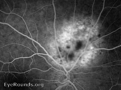
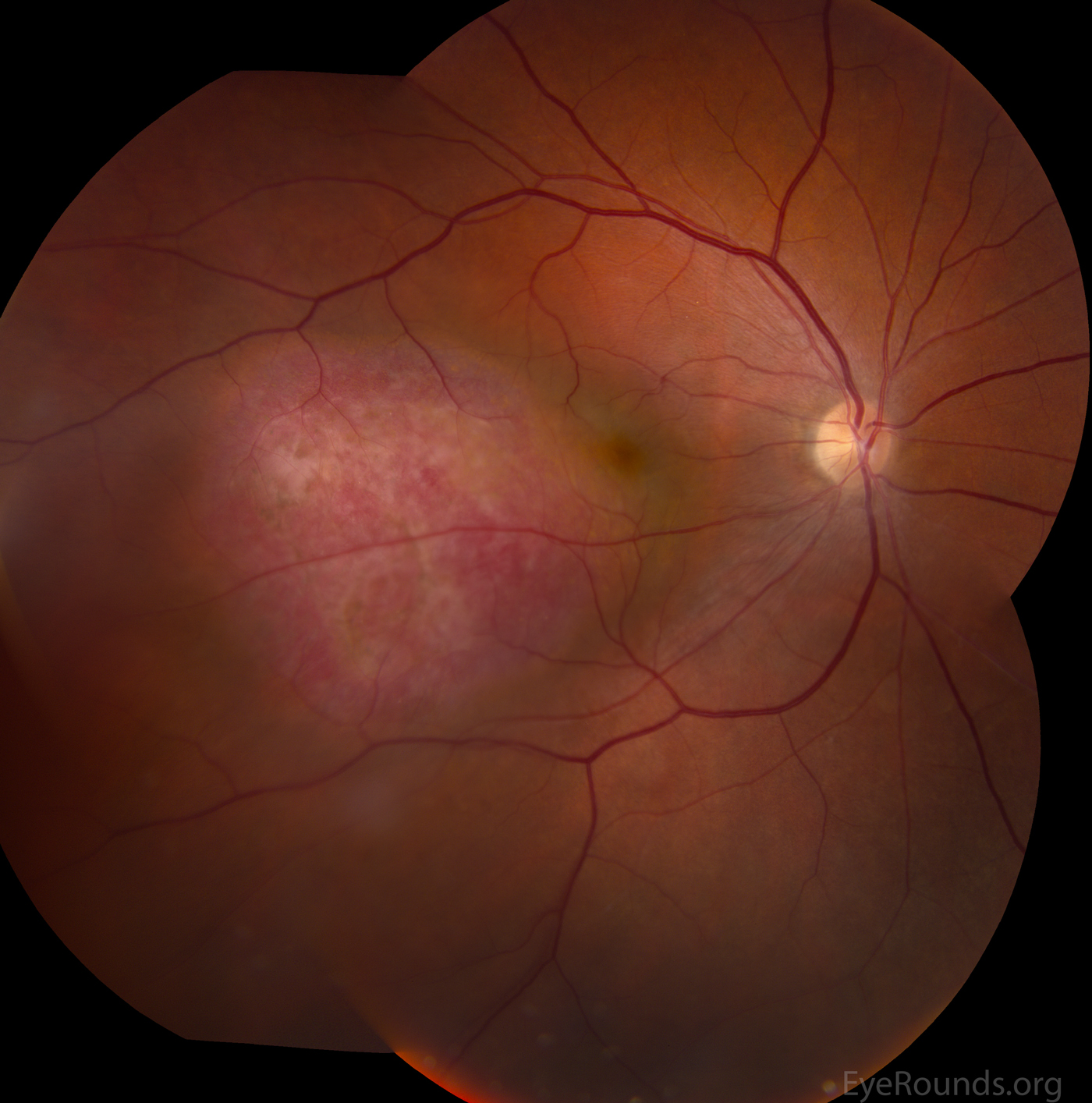
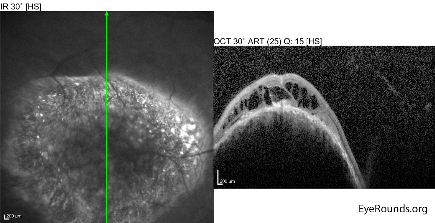
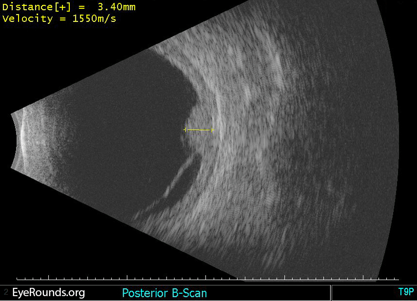
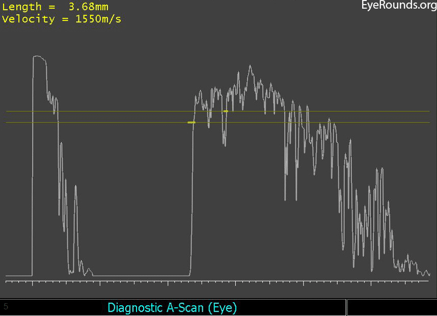
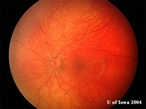
Choroidal hemangiomas are benign vascular hamartomas that can be diffuse (as seen in the case of Sturge-Weber syndrome) or circumscribed as shown here. They typically are red-orange in color with indistinct margins and are located in the posterior pole. There may be overlying retinal pigmented epithelium (RPE) changes or orange pigment. The lesions may have associated intraretinal or subretinal fluid. They display high internal reflectivity on A-scan echography.
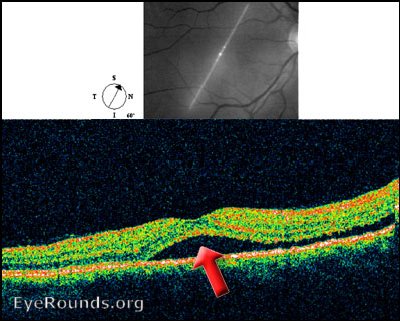
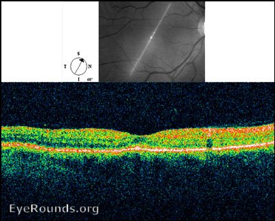
These photographs show the appearance of a circumscribed choroidal hemangioma before and after treatment with photodynamic therapy (PDT). The lesion appeared as an elevated choroidal mass with overlying orange plaques and RPE atrophy. Vascular leakage from the lesion resulted in macular edema overlying the lesion, as seen in the OCT, and surrounding subretinal fluid resulting in an exudative retinal detachment, as seen on the B-scan echography. A-scan echography revealed high internal reflectivity, which is typical for these lesions. Overlying fibrosis can be seen in the post-treatment photograph. The treatment resulted in a reduction in tumor height and decreased subretinal fluid. It was successful in decreasing the patient's metamorphopsia.
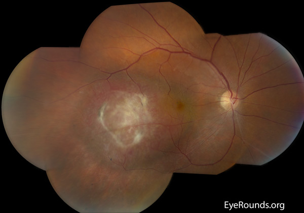
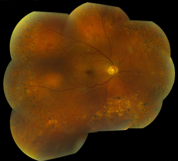
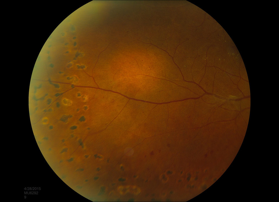
These photographs show the appearance of circumscribed choroidal hemangiomas in two different patients (Figures 2a and 2b are one patient, figures 2c and 2d are another).
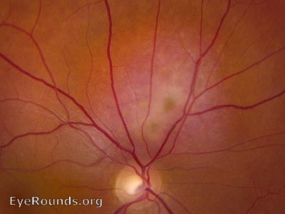
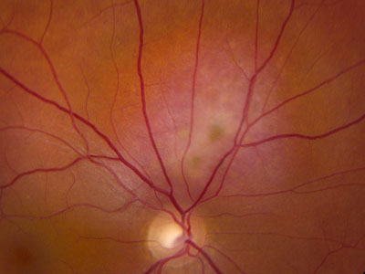
This patient presented with a visually-significant choroidal hemangioma. The patient underwent photodynamic therapy employing the technique of Michels et al. After infusion of 6mg/m2 of verteporfin (Visudyne) intravenously the lesion was illuminated for 163 seconds with the activating laser. Two months later vision had improved from 20/70 to 20/30 and the sub-retinal fluid had resolved clinically and on OCT.
Verteporfin therapy for choroidal hemangioma: a long-term follow-up, Michels, et al Retina. 2005;25(6):697-703.

Ophthalmic Atlas Images by EyeRounds.org, The University of Iowa are licensed under a Creative Commons Attribution-NonCommercial-NoDerivs 3.0 Unported License.

Address
University of IowaLegal
Related Links


