This site uses tracking information. Visit our privacy policy. Click to agree to this policy and not see this again.

Ophthalmology and Visual Sciences

Neuroretinitis

Neuroretinitis

Category(ies): Retina
Contributors: Tyler B.S. Risma, MD; Morgan Bertsch, BS; Jeffrey Welder, MD; Justin Risma, MD; James M. Coombs, MD; Andrew Doan, MD, PhD

Neuroretinitis 1

Neuroretinitis 1

Contributors: Morgan Bertsch, BS and Tyler B.S. Risma, MD

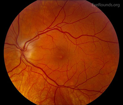
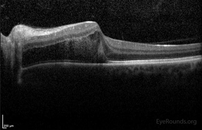
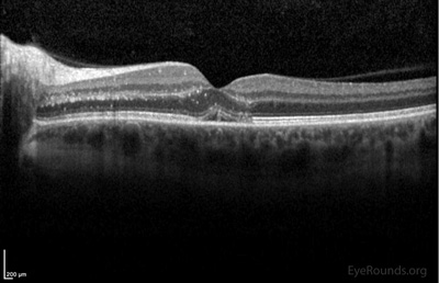
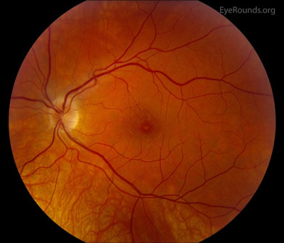
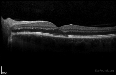
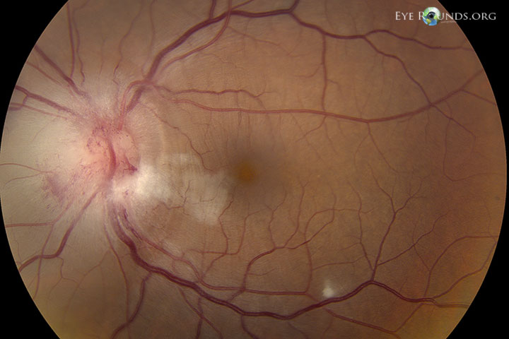
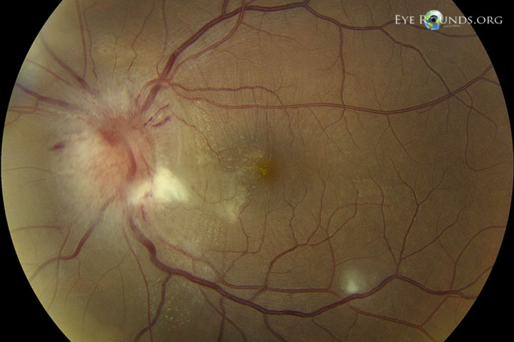
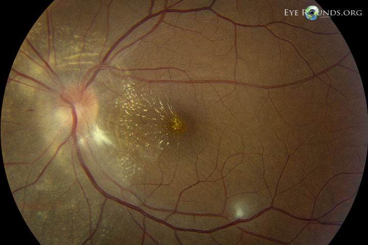
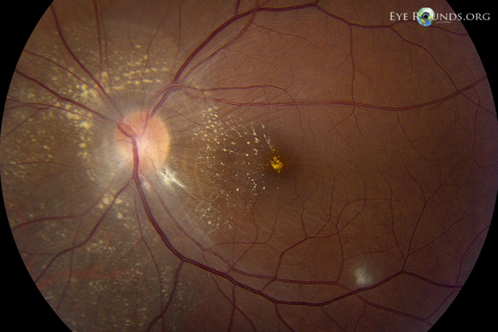
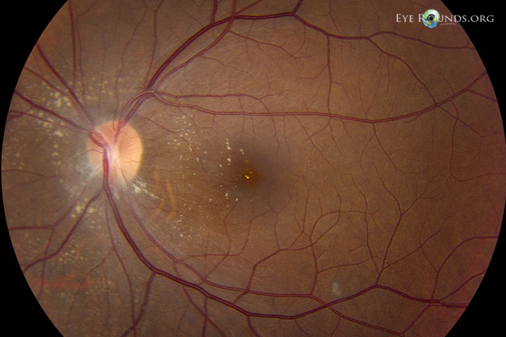
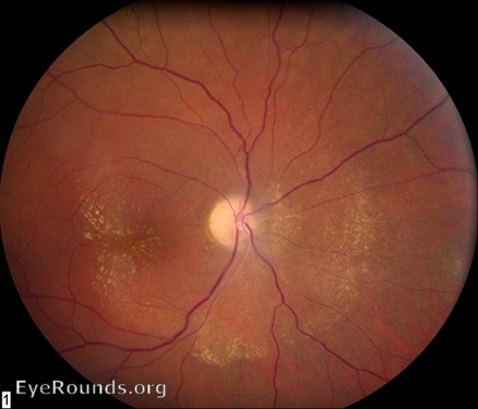
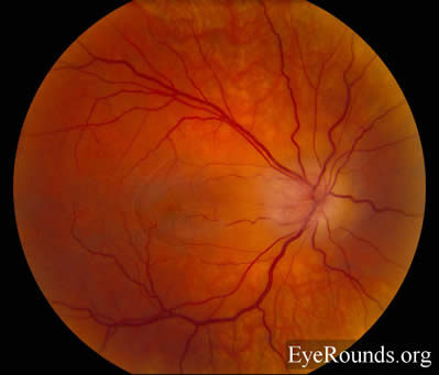
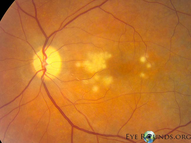
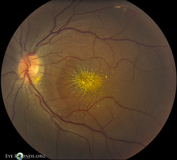
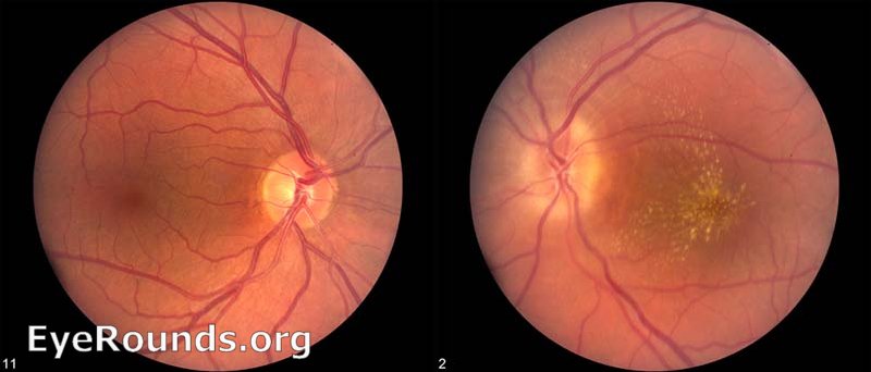
Patient presented with sudden onset decreased vision in his left eye. Fundus photos show the progression of macular star at presentation (Figure 1), 1 week later (Figure 2), and three weeks later (Figure 3). Ocular Coherence Tomography at these same visits shows significant outer retinal fluid in the nasal macula reaching the fovea center. The OCTs show gradual resolution of the intraretinal fluid and deposition of hyper-reflective material in the outer plexiform layer at the fovea, called the nerve fiber layer of Henle, and fluid here gives the classic appearance of a "macular star". The patient was serum positive for Bartonella henselae (Cat Scratch Disease).

Optic Neuroretinitis - fundus day 1
Figure 1a: Fundus Day 1
Optic Neuroretinitis - oct day 1
Figure 1b: Oct Day 1
Optic Neuroretinitis - fundus day 7
Figure 2a: Fundus Day 7
Optic Neuroretinitis - oct day 7
Figure 2b: Oct Day 7
Optic Neuroretinitis - fundus day 21
Figure 3a: Fundus Day 21
Optic Neuroretinitis - oct day 21
Figure 3b: Oct Day 21

Neuroretinitis 2: Bartonella Neuroretinitis

Neuroretinitis 2: Bartonella Neuroretinitis1

Contributor: Jeffrey Welder, MD

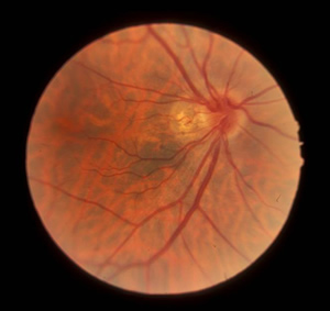
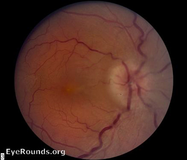
34-year-old female diagnosed with Bartonella neuroretinitis and cilioretinal artery occlusion of the left eye. She presented with a "gray spot" in her vision. Exam revealed an RAPD OS. Photos demonstrate appearance on presentation, 1 week, 3 weeks, 4 weeks, and 8 weeks after presentation. Patient was treated with oral Azithromycin at 5 weeks after presentation when positive bartonella serology returned.

Photo sequence:
Optic Neuroretinitis Bartonella neuroretiniti Initial presentation
Initial presentation

Neuroretinitis 3

Neuroretinitis 3

Contributor: Justin Risma, MD
Neuroretinitis 3 fundus
17-year-old male with painless central vision loss in the left eye. Dilated exam revealed disc edema and a macular star (hard exudates arranged in a radial pattern due to the alignment of fibers in the outer plexiform layer, or Henle's layer).

Neuroretinitis 4

Neuroretinitis 4

Contributor: James M. Coombs, MD
Neuroretinitis Idiopathic
43-year-old female with acute loss of vision in the right eye. Infectious/inflammatory work-up negative
Neuroretinitis Idiopathic 2 months after steroids
2 months later after a course of oral steroids

Neuroretinitis 5

Neuroretinitis 5

Contributor: Andrew Doan, MD, PhD
Neuroretinitis disc swelling with macular star
Patient with unilateral vision loss in the left eye. Marked disc swelling with macular star figure. Serum positive for bartonella henselae (Cat Scratch Disease).