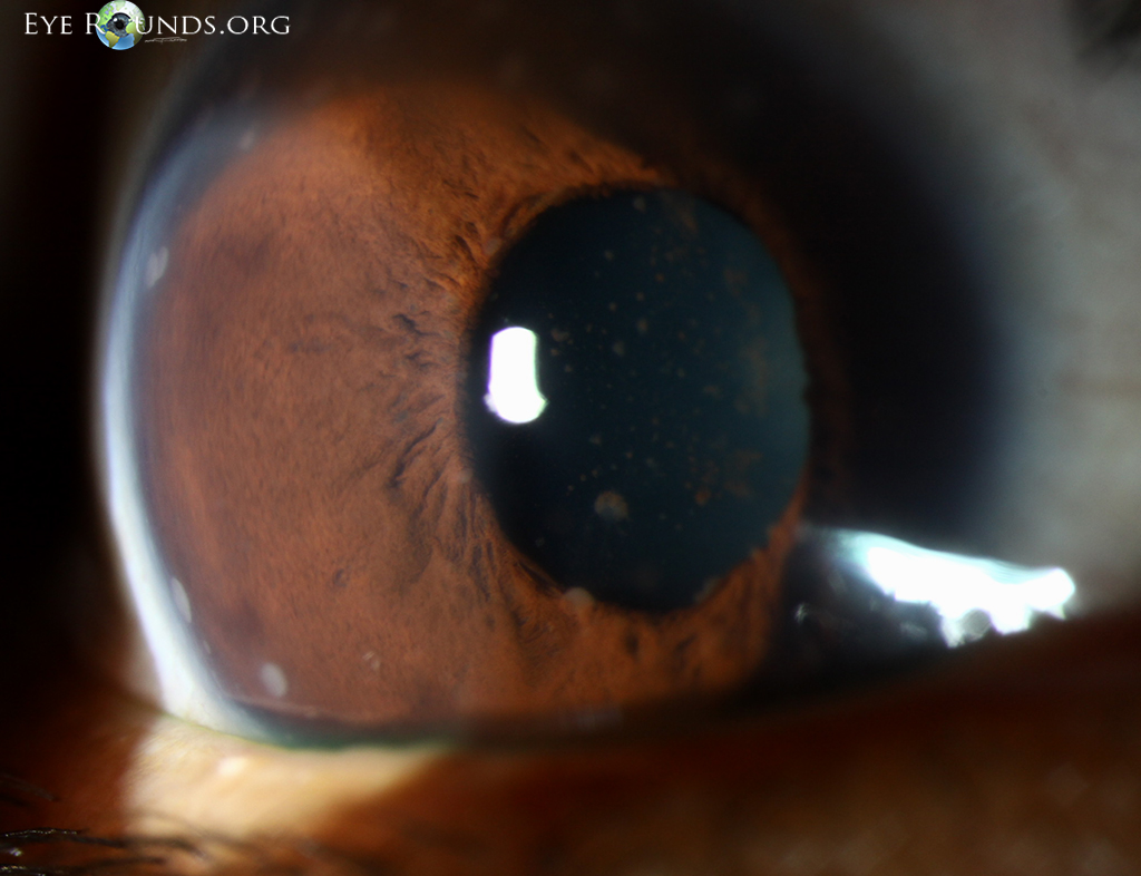
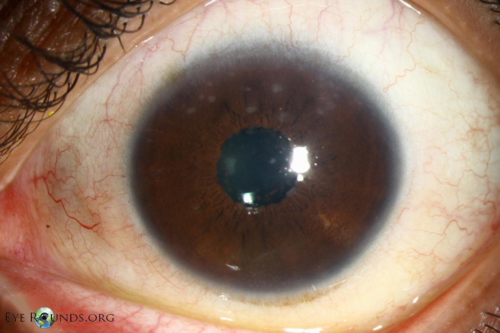
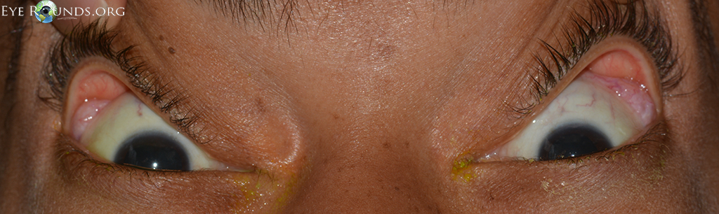
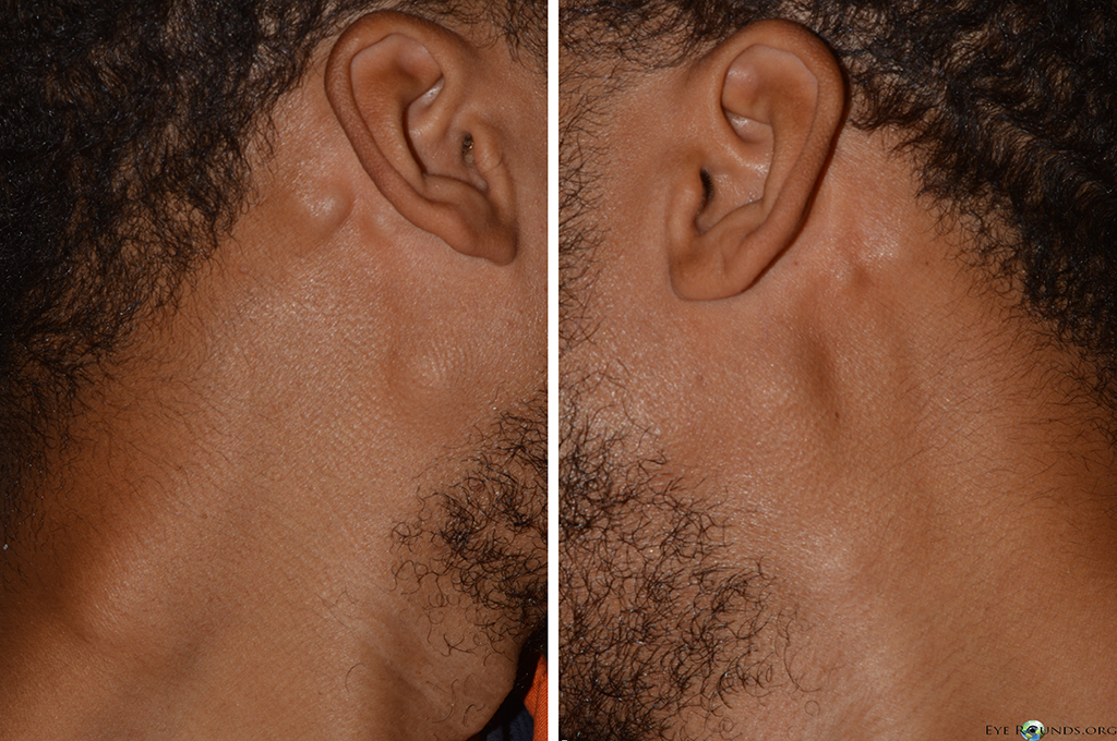
This 34-year-old African American male presented with a few months of blurry vision and photophobia. On exam, he was noted to have bilateral granulomatous anterior uveitis with large mutton-fat keratic precipitates, iris granulomas, enlarged lacrimal glands, and bilateral cervical and post-auricular lymphadenopathy. Laboratory testing revealed an angiotensin converting enzyme (ACE) level of 294 U/L (normal 8-52). He was referred to pulmonary medicine and diagnosed with sarcoidosis.

Ophthalmic Atlas Images by EyeRounds.org, The University of Iowa are licensed under a Creative Commons Attribution-NonCommercial-NoDerivs 3.0 Unported License.


Address
University of IowaLegal
Related Links