

EyeRounds Online Atlas of Ophthalmology
Contributor: William Charles Caccamise, Sr, MD, Retired Clinical Assistant Professor of Ophthalmology, University of Rochester School of Medicine and Dentistry
*Dr. Caccamise has very generously shared his images of patients taken while operating during the "eye season" in rural India as well as those from his private practice during the 1960's and 1970's. Many of his images are significant for their historical perspective and for techniques and conditions seen in settings in undeveloped areas.
Category: Systemic Disorders
Vitamin A deficiency with xerosis conjunctivae and a nasal Bitot's spot
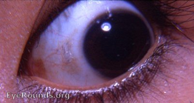
Some textbooks state that Bitot's spots can occur on either side of the cornea. Dr. Caccamise's experience with innumerable cases of vitamin A deficiency was as follows: In most cases the more common temporal Bitot's spot was not accompanied by a nasal Bitot's spot. When a nasal Bitot's spot was present, there was invariably a more developed temporal Bitot's spot.
In the field in a third world atmosphere, a Bitot's spot means vitamin A deficiency until proved otherwise. The xerotic conjunctival area that forms the floor of the Bitot's spot in the photo is evident as the discolored bulbar conjunctival area nasal to the limbus. An epicanthal fold is evident nasally.

Ophthalmic Atlas Images by EyeRounds.org, The University of Iowa are licensed under a Creative Commons Attribution-NonCommercial-NoDerivs 3.0 Unported License.