Acute Angle Closure Glaucoma
Acute Angle Closure Glaucoma
Category(ies): Glaucoma/Iris
Posted: February 8, 2008
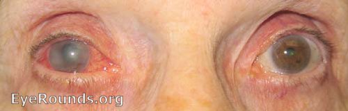
- Patient awoke with severe pain OD, headache, decreased vision OD, and nausea. Photo shows unilateral redeye with hazy cornea. Right eye was very firm with a pressure of 70+ mmHg. Refractive error was +1.50 to +2.00 sphere in each eye.
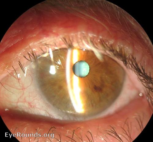
- Examination of the left eye showed a narrow anterior chamber by comparing the width between slit beams.
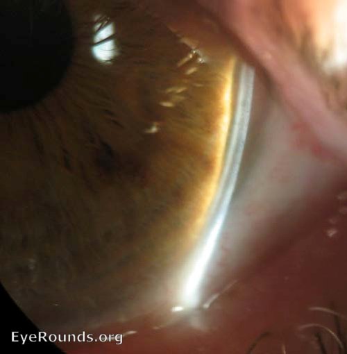
- Von Herrick Test showed a narrow angle in the left eye. Distance between slit beams should be at least 1/2 of the corneal thickness (~250 microns). Gonioscopy demonstrated a closed angle OS too.
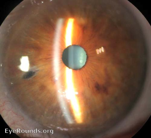
- Laser peripheral iridotomies (LPI) were performed on both eyes. Aqueous suppressants were started OD and the pressure dropped.