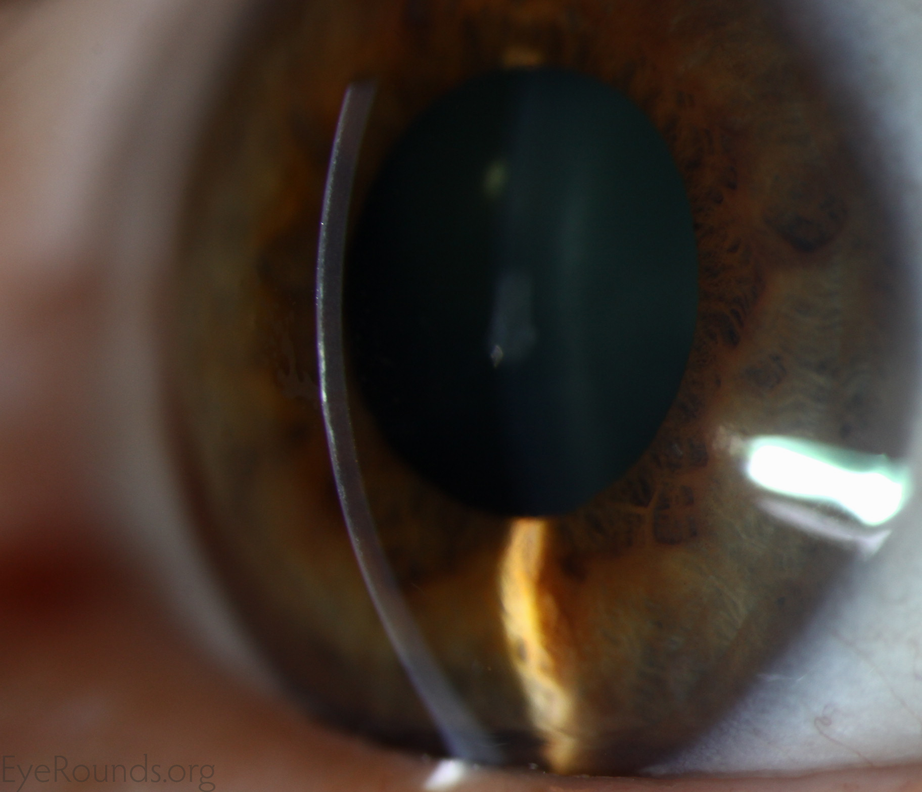
Epithelial ingrowth occurs when the corneal epithelial cells grow within the lamellar interface beneath the LASIK flap. This may result in an inflammatory response, corneal edema, irregular astigmatism, or melting of the LASIK flap. When mild and peripheral, it may be observed. When progressive or severe, the LASIK flap must be lifted and the epithelium scraped from the bed. Recurrent cases may require suturing of the LASIK flap.
Contributor: Jesse Vislisel, MD
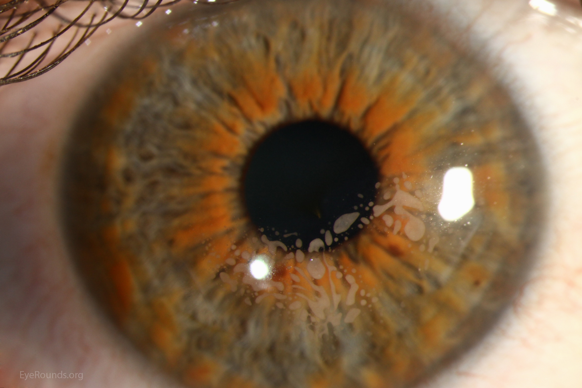
Nests of epithelium can be seen approaching the visual axis beneath the LASIK flap. This case occured after microkeratome-assisted LASIK and multiple enhancement procedures
Contributor: Jesse Vislisel, MD
Photographer: Brice Critser, CRA
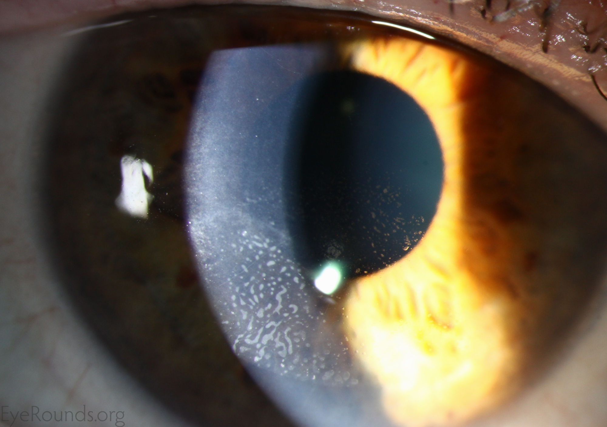
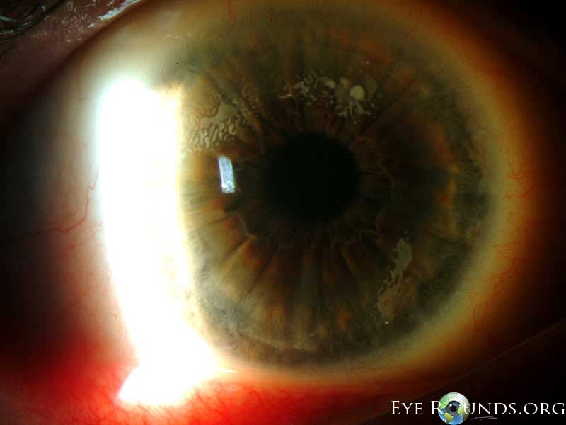
Epithelial ingrowth after LASIK performed in the setting of epithelial basement membrane dystrophy. The patient developed edema of the LASIK flap and recurrent erosion syndrome, requiring lifting of the flap and removal of the epithelial ingrowth
Contributor: Jesse Vislisel, MD
Photographer: Brice Critser, CRA
Contributor: Matt Ward, MD
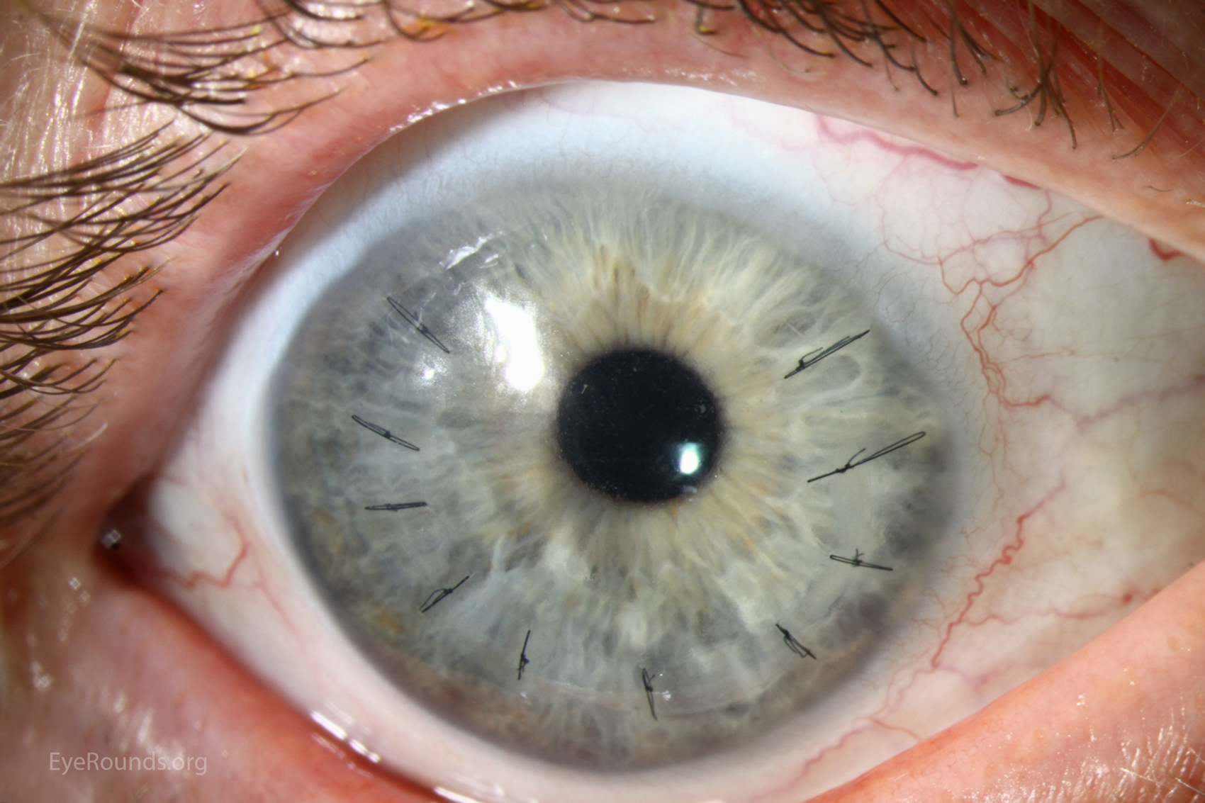
Epithelial ingrowth can be a vision threatening complication of LASIK surgery. If it approaches the visual axis, the flap must be lifted and the bed scraped. Recurrent ingrowth may require suturing of the flap in the location of recurrence.

Ophthalmic Atlas Images by EyeRounds.org, The University of Iowa are licensed under a Creative Commons Attribution-NonCommercial-NoDerivs 3.0 Unported License.

Address
University of IowaLegal
Related Links