The facial colliculus is an anatomic elevation on the floor of the 4th ventricle located medial to the sulcus limitance in the dorsal pons. It is formed by the abducens nucleus (CN VI) and the fascicle of the facial nerve (CN VII) as the motor fibers of CN VII loop around the CN VI nucleus (internal genu of the facial nerve). Patients with a facial colliculus syndrome will demonstrate an ipsilateral horizontal gaze palsy with an ipsilateral lower motor neuron pattern of facial weakness. The horizontal gaze palsy is secondary to a lesion that is affecting the CN VI nucleus that is responsible for abduction of the ipsilateral eye by sending projections to the ipsilateral lateral rectus muscle, but is also responsible for initiating conjugate adduction of the contralateral eye by sending projections the contralateral medial longitudinal fasciculus (MLF) that signal the medial rectus subnucleus of cranial nerve III (CN III) that projects to the medial rectus of the contralateral eye. The ipsilateral lower motor neuron pattern of facial weakness is caused by disruption of the fascicle of CN VII.
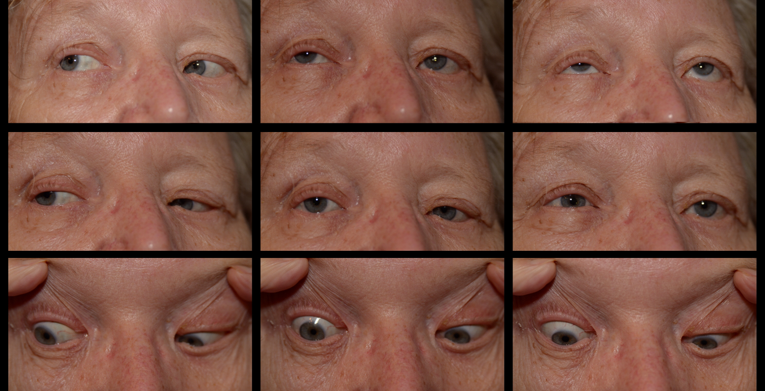
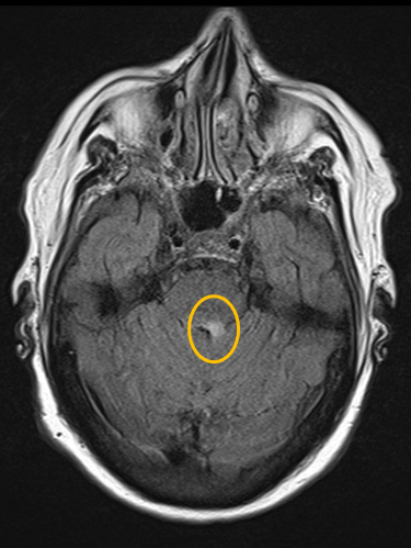
This patient presented with complaints of diplopia and left facial drooping. She was noted to have an esotropia in primary gaze that resolved in right gaze and adopted a compensatory right face turn. She had a left horizontal gaze palsy that could not be overcome with vestibulo-ocular reflex. She had dense facial weakness on the left (loss of forehead wrinkling, lower eyelid ectropion, poor blink, brow ptosis, flattened nasolabial fold, and a left mouth droop). Imaging showed an enhancing lesion in the dorsal left pons consistent with her diagnosis of CNS lymphoma.

Ophthalmic Atlas Images by EyeRounds.org, The University of Iowa are licensed under a Creative Commons Attribution-NonCommercial-NoDerivs 3.0 Unported License.

Address
University of IowaLegal
Related Links