This site uses tracking information. Visit our privacy policy. Click to agree to this policy and not see this again.

Ophthalmology and Visual Sciences

Corneal melt around Boston type I keratoprosthesis

Corneal melt around Boston type I keratoprosthesis

Category(ies): Cornea, External Disease
Contributor: Jesse Vislisel, MD

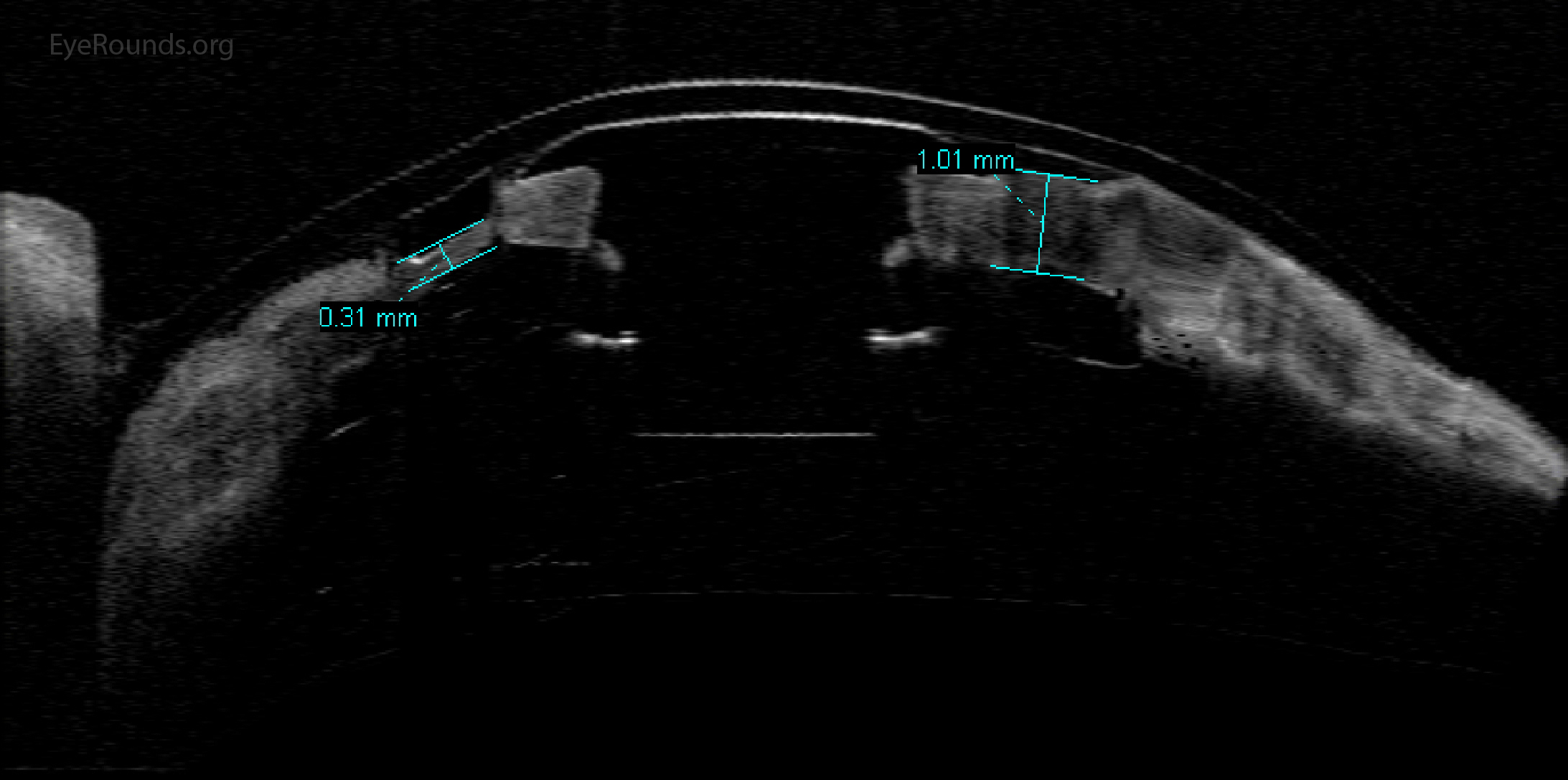
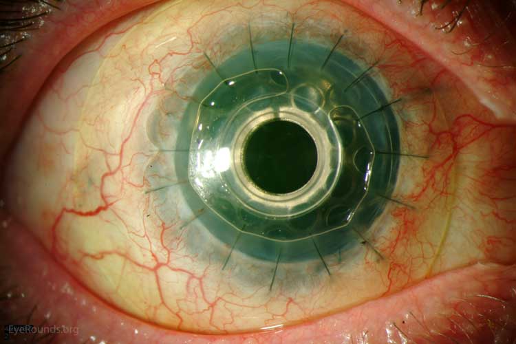
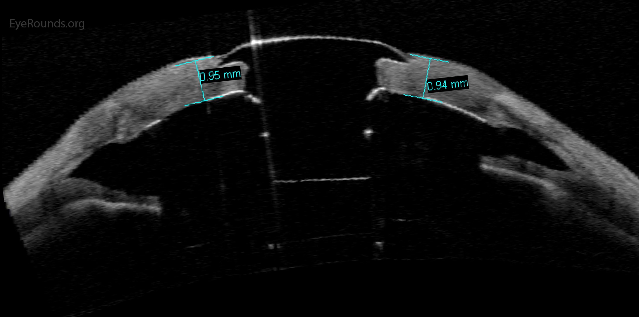
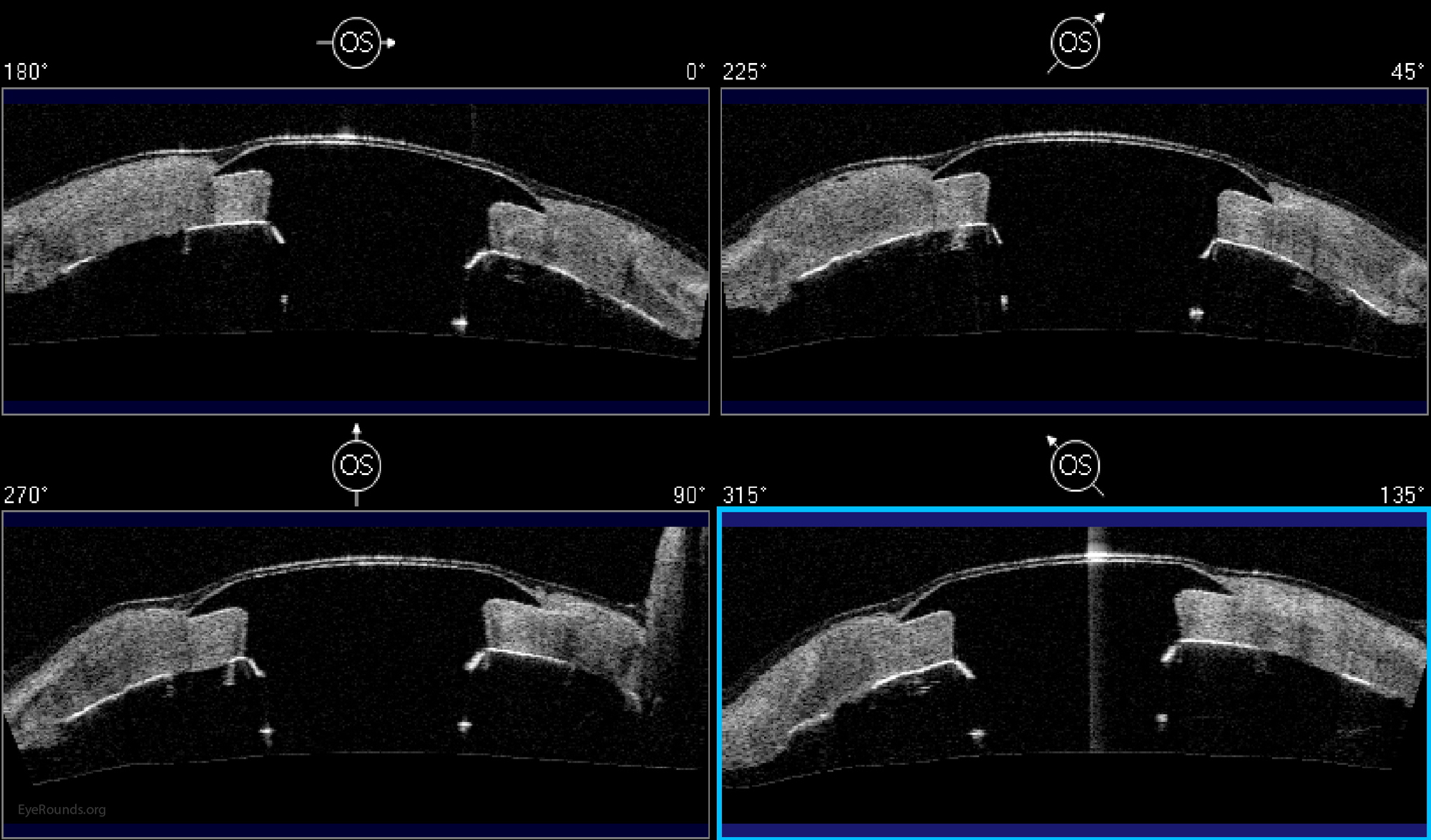
Boston keratoprosthesis devices require regular monitoring to assess for the presence of complications such as infection and corneal melt. Anterior segment optical coherence tomography (OCT) is a useful study to assess for the presence of corneal thinning or melt. We regularly use OCT to monitor the status of the cornea around the device.

Boston type I keratoprosthesis device on anterior segment OCT
Figure 1: Normal appearance of Boston type I keratoprosthesis device on anterior segment OCT.
Boston type I keratoprosthesis device on anterior segment OCT with healthy corneal tissue
Figure 2: Anterior segment optical coherence tomography demonstrating the appearance of a Boston Type I Kpro with healthy corneal tissue surrounding the device
Corneal melt with thinning surrounding the device stem
Figure 3: Corneal melt with thinning of the tissue surrounding the device stem.
Corneal melt with tissue retraction away from the device stem (arrow)
Figure 4: Corneal melt with tissue retraction away from the device stem (arrow).