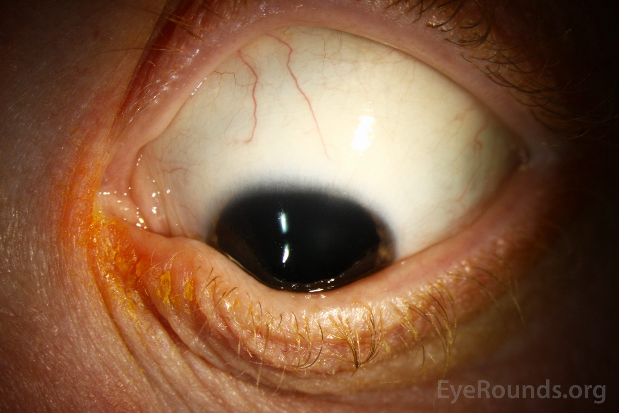
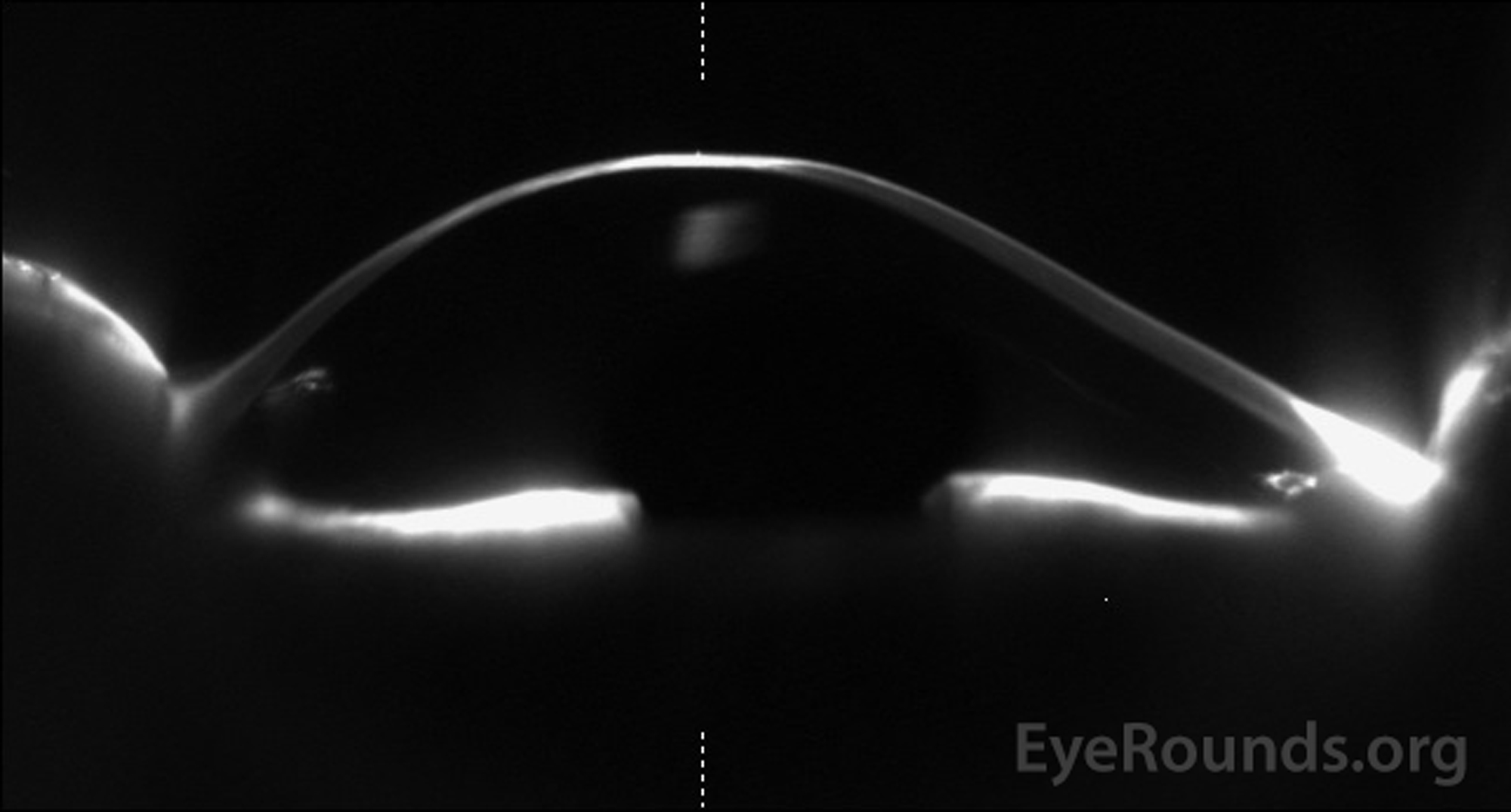
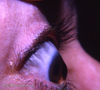
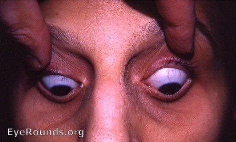
Figure 2. Slit lamp photograph of the left eye of a patient with keratoconus shows the cone-shaped cornea creating an indentation or bowing of the lower eyelid in downgaze, also known as Munson's sign.
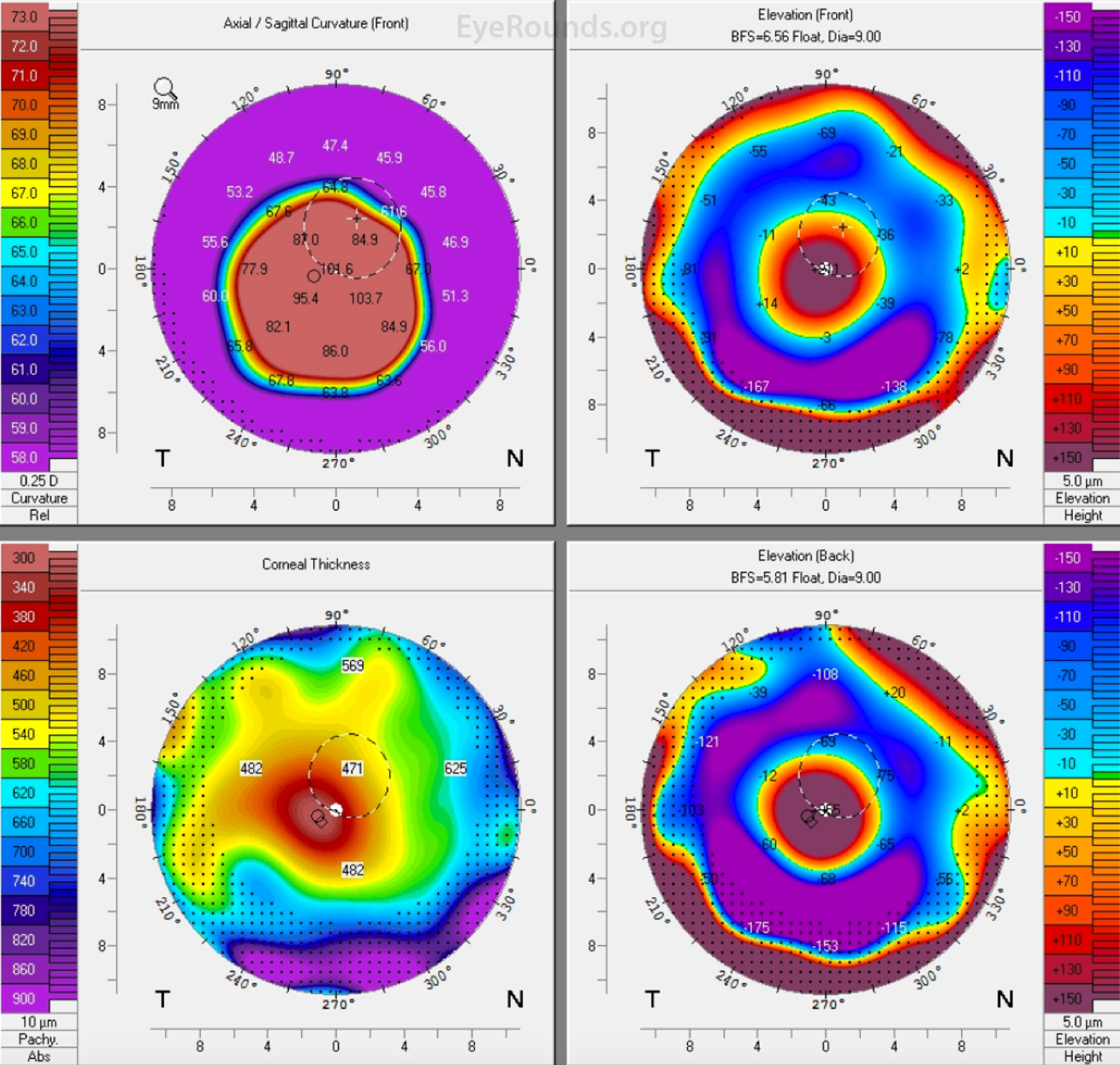
Figure 4. Pentacam 4 Map Report of a patient with advanced keratoconus. The axial curvature map depicts a steep anterior corneal curvature centrally with a large difference in refractive power at the apex compared to the periphery. The anterior float and posterior float maps also depict the elevated central cornea compared to the depressed periphery. The corneal thickness map depicts advanced central corneal thinning, consistent with advanced keratoconus.
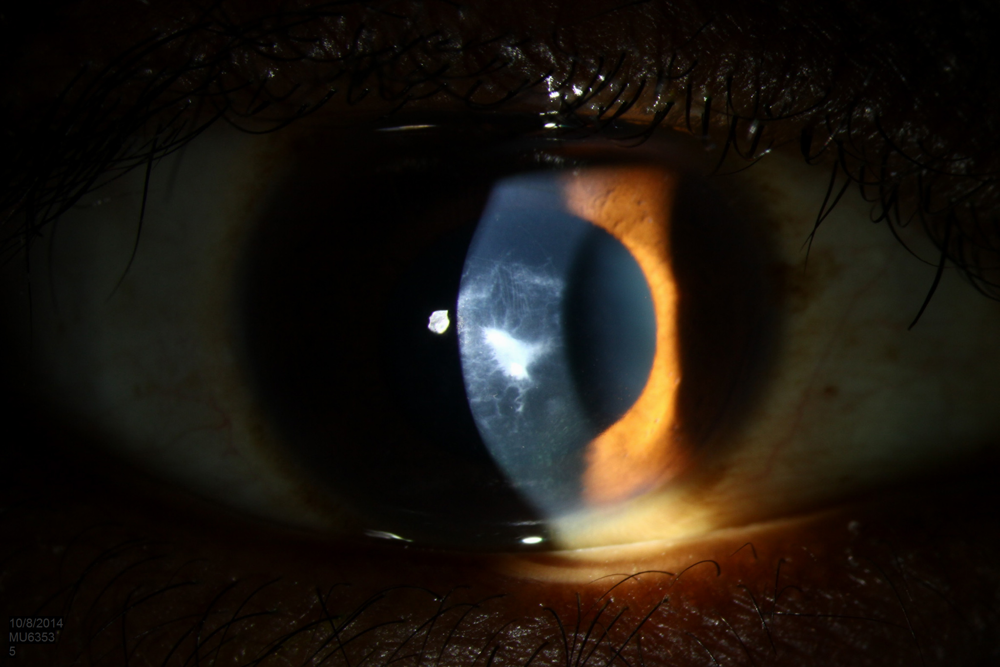
Keratoconus is a bilateral corneal ectasia characterized by central thinning and bulging of the cornea resulting in a cone-shaped protrusion. This patient demonstrates multiple characteristic signs of the condition.
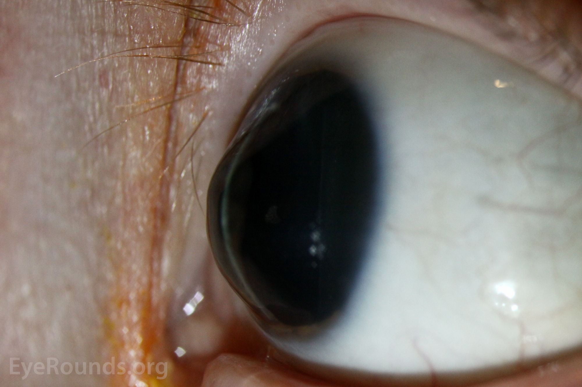
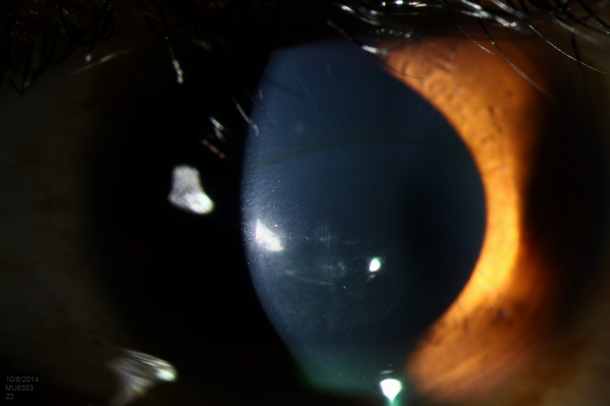
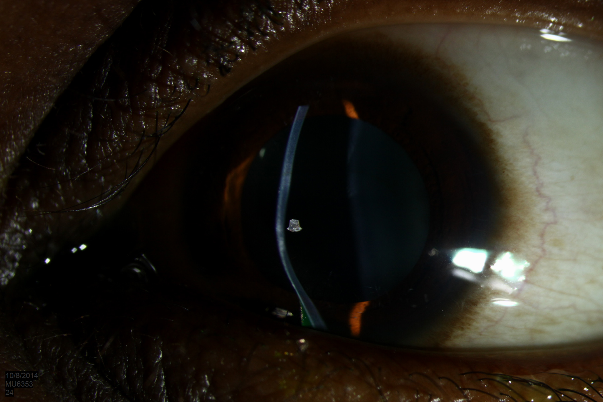
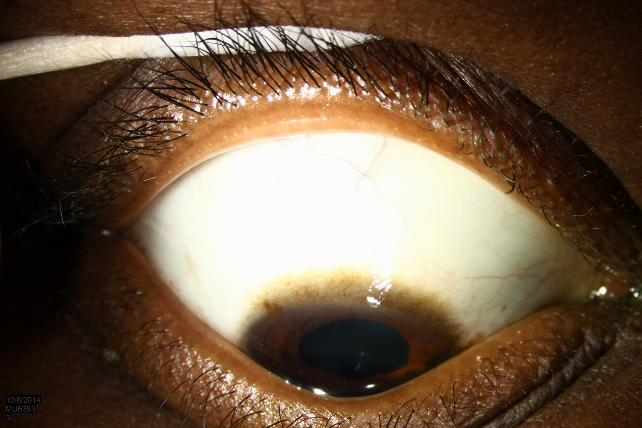
Figure 1. Demonstrates central Vogt's striae, or parallel, vertical lines from posterior stromal stress. A Fleischer ring, or epithelial iron deposition around the base of the cone is visible superiorly. The horizontally-oriented opacities are areas of stromal scarring.
Contributor: William Charles Caccamise, Sr, MD,Retired Clinical Assistant Professor of Ophthalmology, University of Rochester School of Medicine and Dentistry
*Dr. Caccamise has very generously shared his images of patients taken while operating during the "eye season" in rural India as well as those from his private practice during the 1960's and 1970's. Many of his images are significant for their historical perspective and for techniques and conditions seen in settings in undeveloped areas.
Figure 1. The photo shows scarring of the cornea at the apex of the keratoconus.
Figure 2. This female patient demonstrated the typical findings with keratoconus: There was conical ectasia (bulging) with an irregular myopic astigmatism. Keratoconus is sometimes called ectatic corneal dystrophy. 70% of the cases occur in females.
University of Iowa
Roy J. and Lucille A. Carver College of Medicine
Department of Ophthalmology and Visual Sciences
200 Hawkins Drive
Iowa City, IA 52242
University of Iowa
Roy J. and Lucille A. Carver College of Medicine
Department of Ophthalmology and Visual Sciences
200 Hawkins Drive
Iowa City, IA 52242