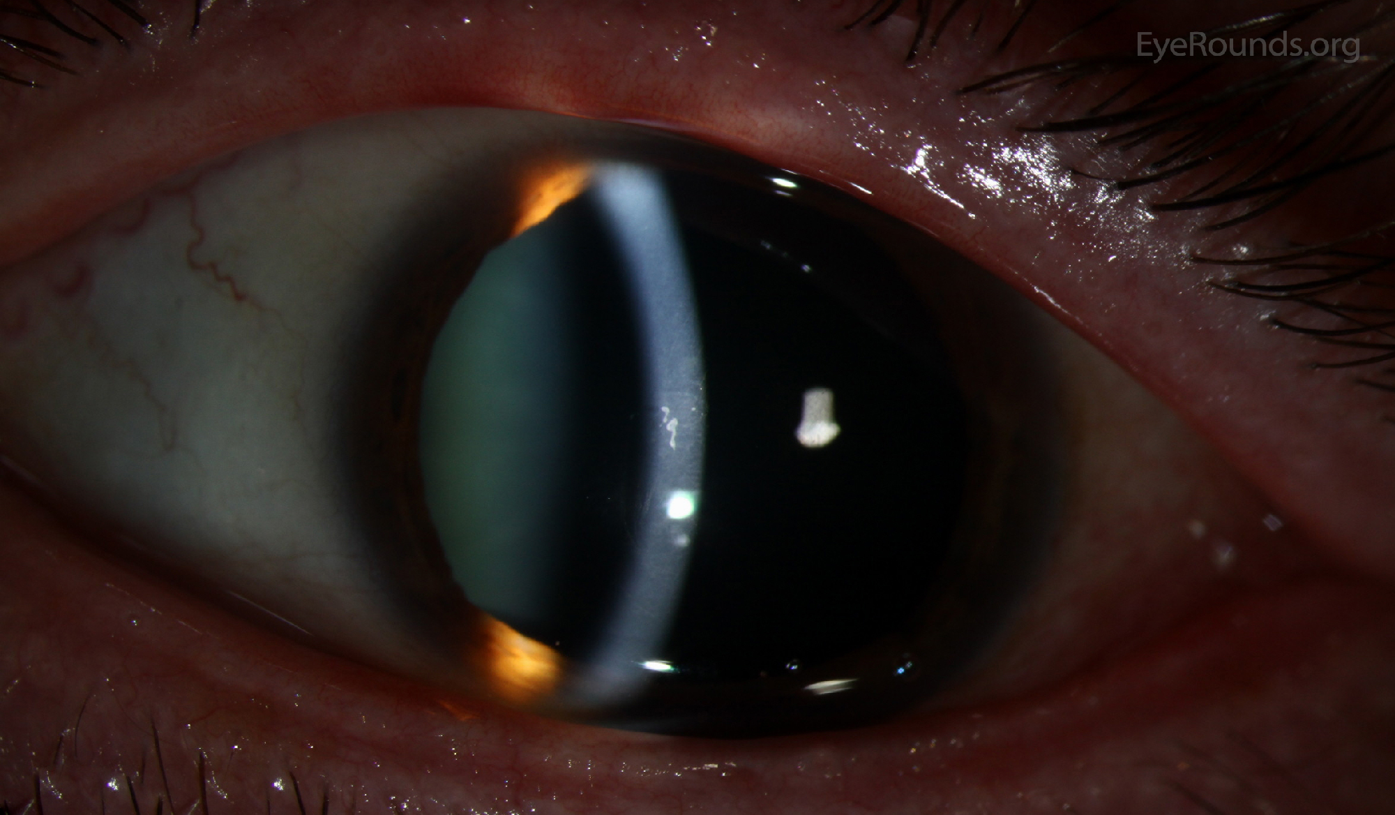
This patient had both Meesmann epithelial corneal dystrophy and striking epithelial basement membrane dystrophy (EBMD), neither of which were symptomatic.
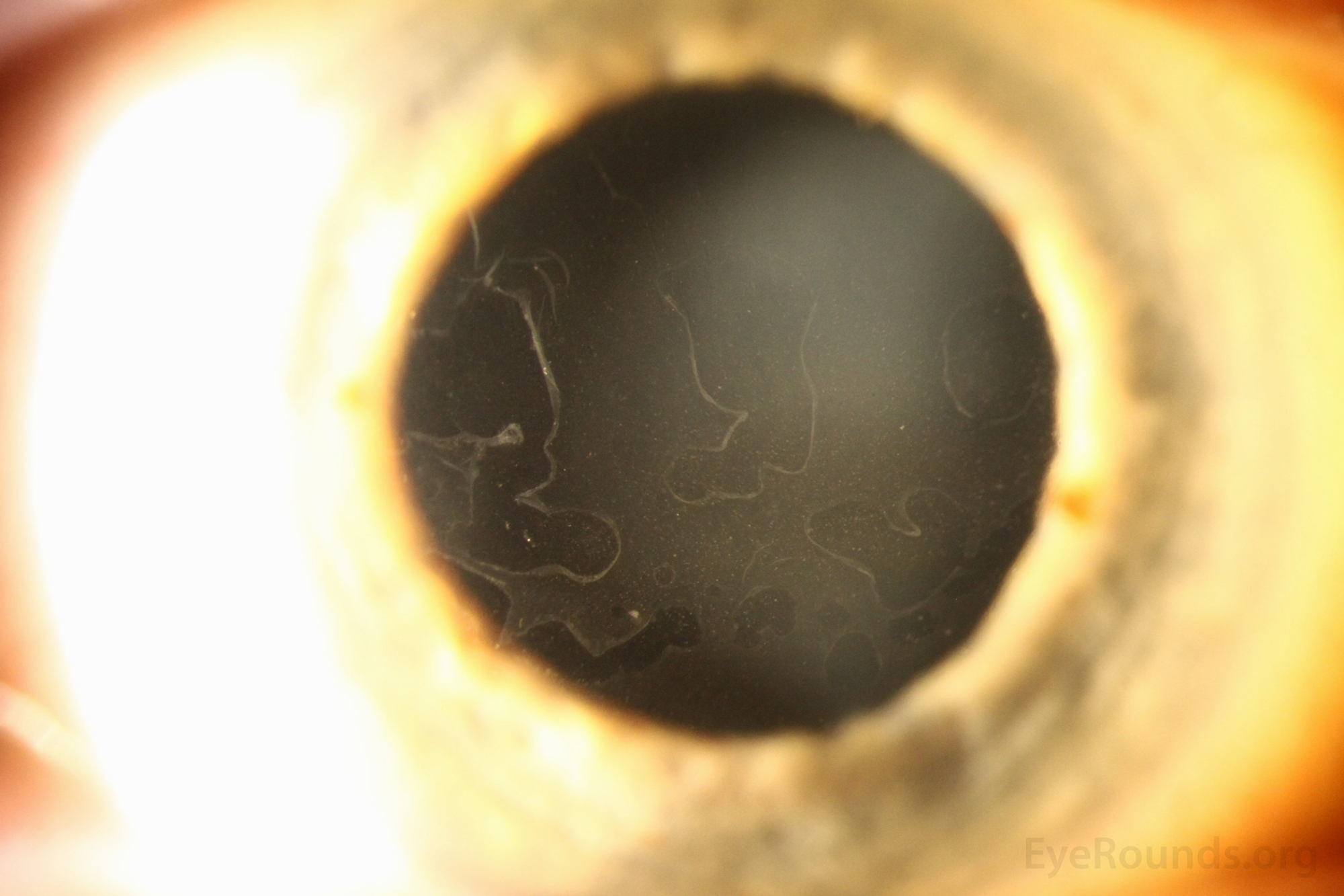
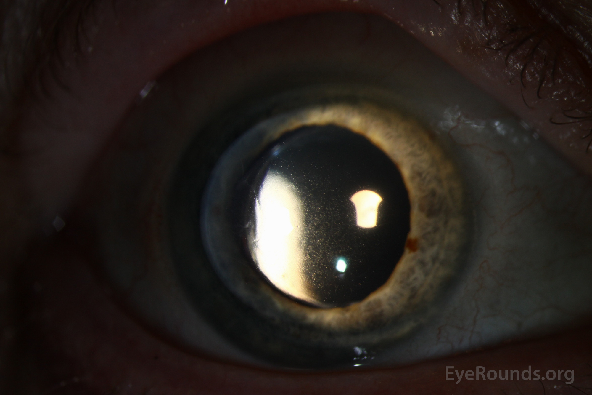
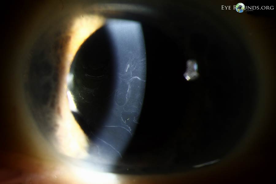
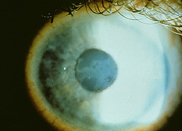
72-year-old female with decrease in central visual acuity. Classic gray patches, fingerprint lines, map lines, dots, and cobblestone pattern in the corneal epithelium are seen on this photo.
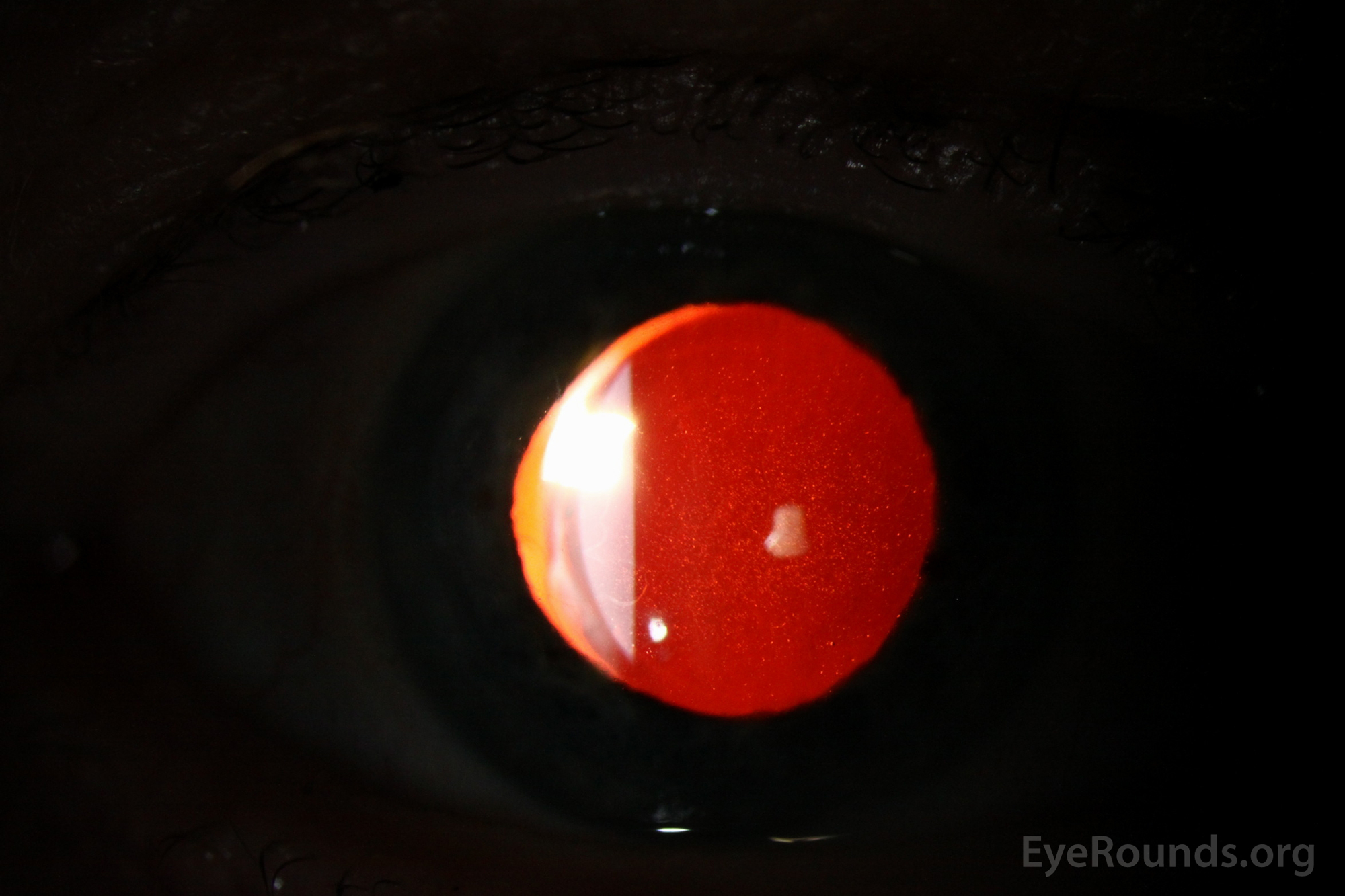
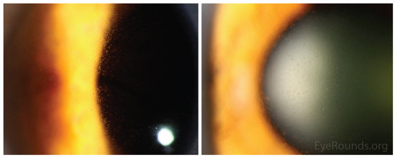
This photo demonstrates classic "dots", which manifest as irregular round, oval, or comma-shaped, non-staining, putty-gray intraepithelial opacities. They represent intraepithelial pseudocysts containing cytoplasmic debris.
Weiss JS, Moller HU, Aldave AJ, et al. IC3D Classification of Corneal Dystrophies—Edition 2. Cornea. 2015;34:117-159.

Ophthalmic Atlas Images by EyeRounds.org, The University of Iowa are licensed under a Creative Commons Attribution-NonCommercial-NoDerivs 3.0 Unported License.

Address
University of IowaLegal
Related Links