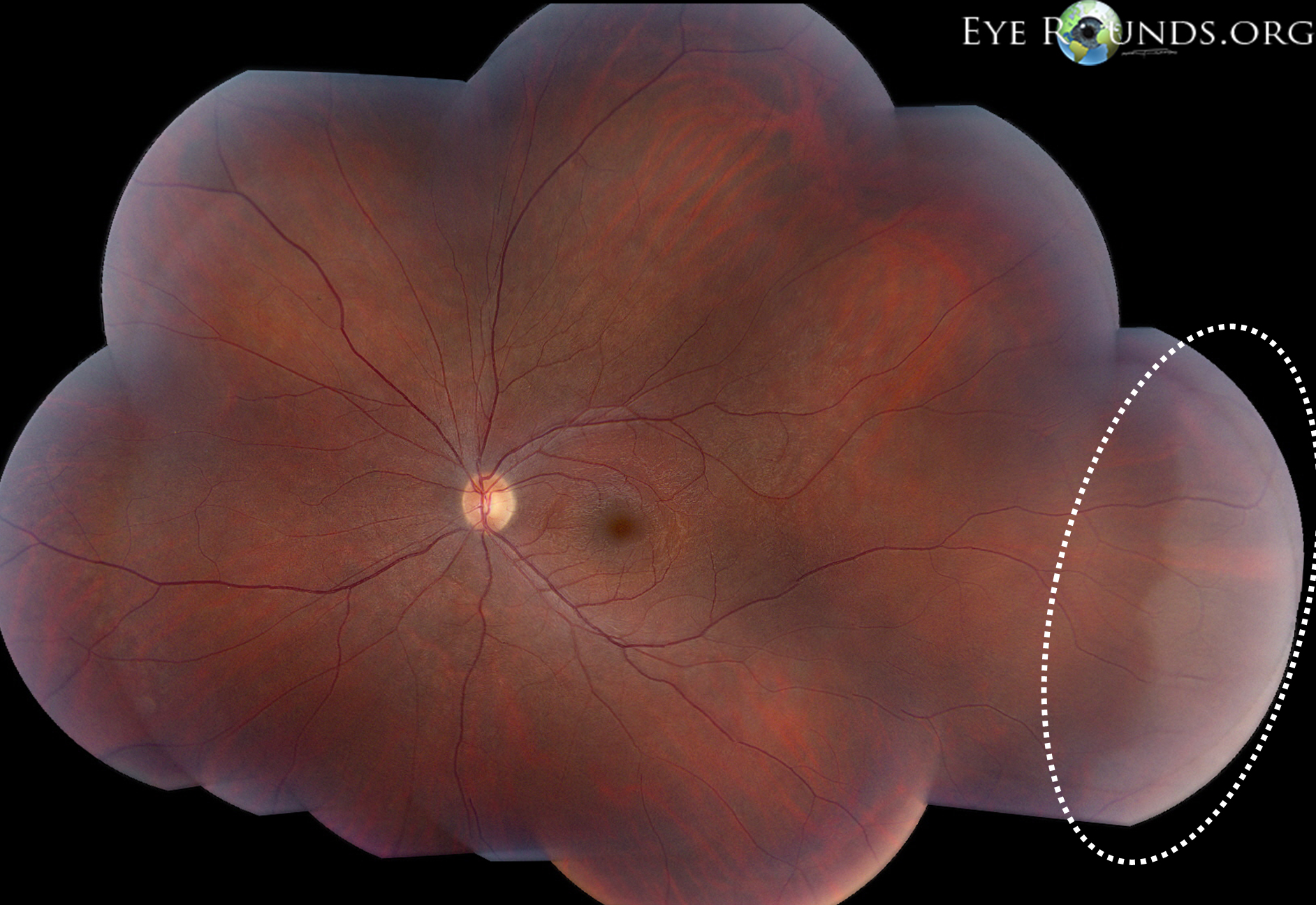
A 20-year-old male was seen in the Retina Clinic for the possibility of retinitis pigmentosa, both eyes. Unrelated to this, the peripheral exam showed relatively symmetric white patches of retina inferiorly and inferotemporally that were visible without scleral indentation. Scleral depressed exam revealed that the retina was attached, and that there was no other peripheral retinal pathology. The vision and remainder of the exam was otherwise unremarkable.
The exact cause of WWP is unknown. There has been controversy regarding its anatomical location, associated pathology, and its potential association with the development of retinal breaks.[1]
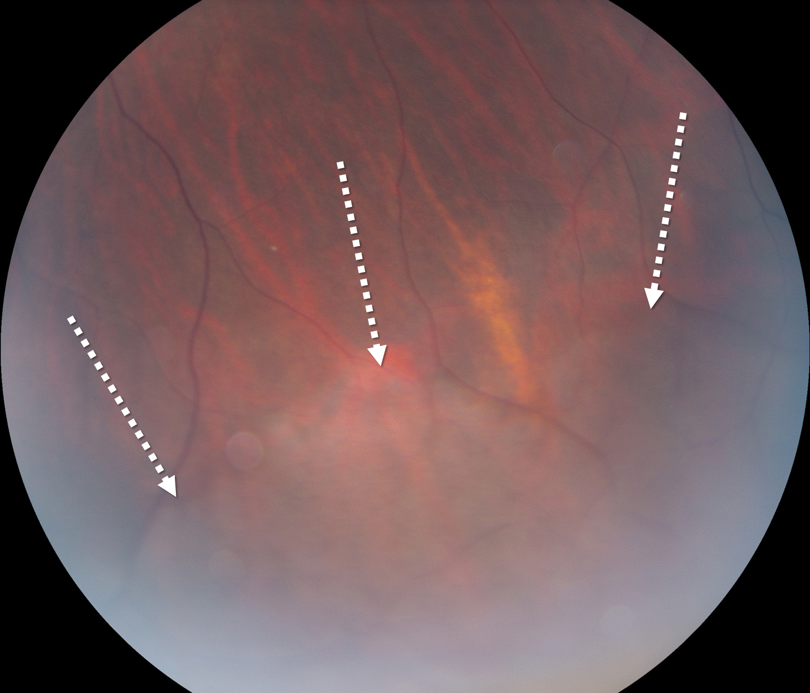
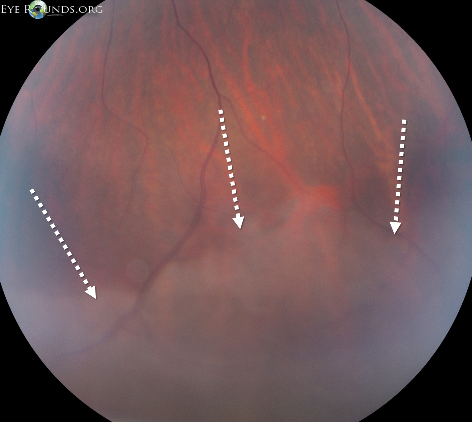
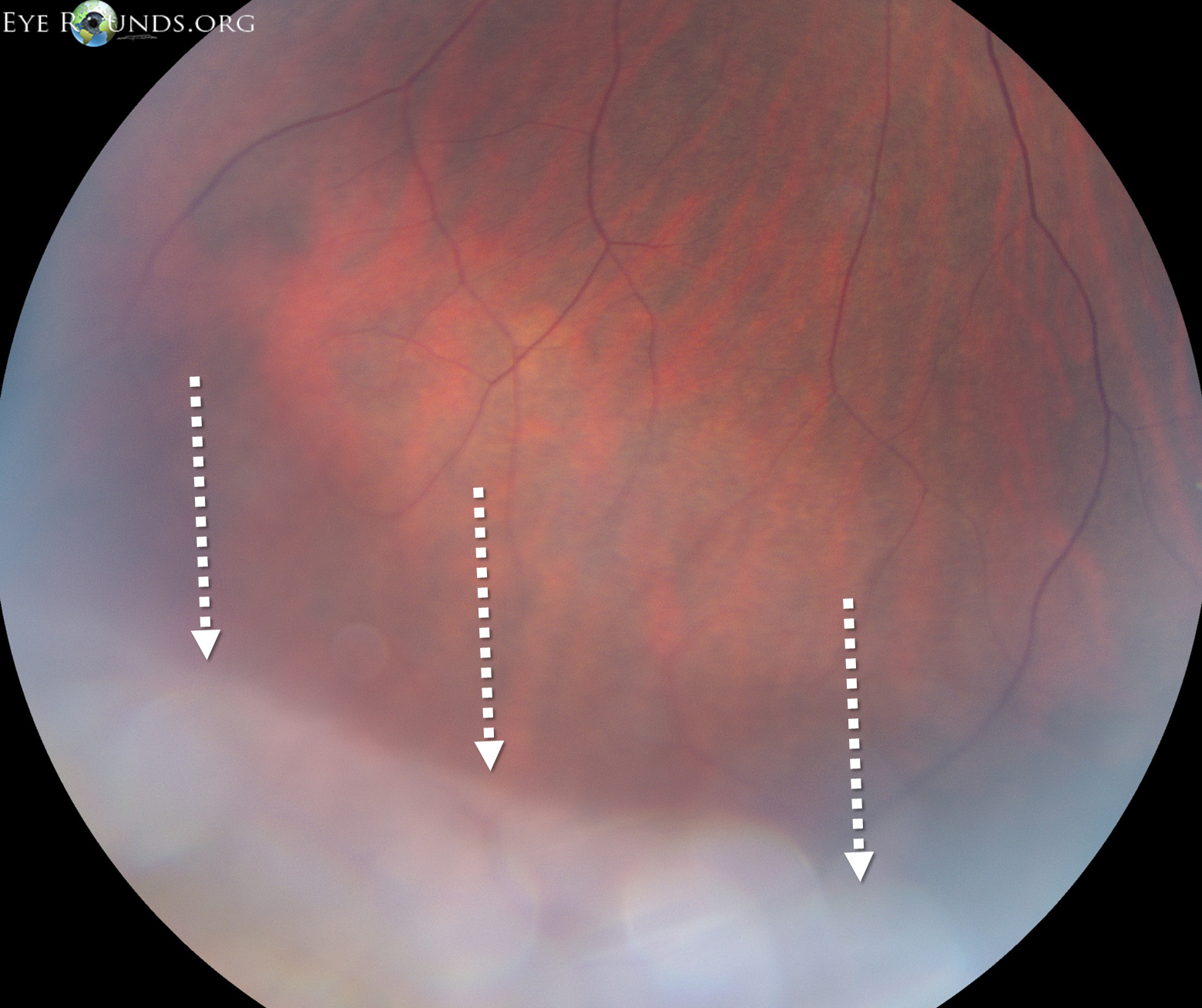
Clinically, a white appearance of the equator and/or peripheral retina seen without scleral indentation. Margins are often sharply demarcated from normal retina.
Frequently misdiagnosed as a retinal detachment or retinal schisis; however, scleral indentation reveals that the retina is apposed to the underlying RPE.
Reports show that OCT corresponds to a distinct hyper-reflectance of the outer retina, likely the elipsoid zone on SD-OCT.[2]

Ophthalmic Atlas Images by EyeRounds.org, The University of Iowa are licensed under a Creative Commons Attribution-NonCommercial-NoDerivs 3.0 Unported License.

Address
University of IowaLegal
Related Links