Chief Complaint: 31-year-old caucasian female presented with a painless, "dark spot" in her left visual field.
History of Present Illness: Dark spot fixed in supero-nasal left visual field that she noticed for one day. No complaints of flashes or floaters. Patient noted that the dark spot in her vision clears with abduction.
Past Ocular History: Peripheral atrophic holes OD
Medical History: Cerebral palsy
Social History: Librarian. No tobacco or alcohol.
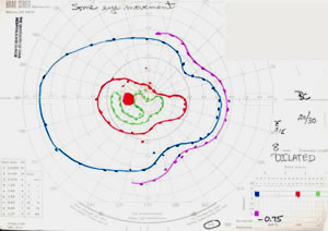
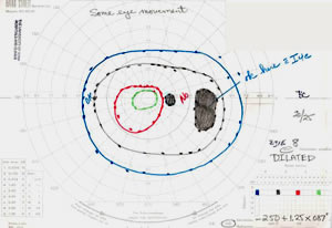
| GVF OS with supero-nasal and infero-nasal constriction of all isopters. | GVF OD with temporal scotoma. |
 |
 |
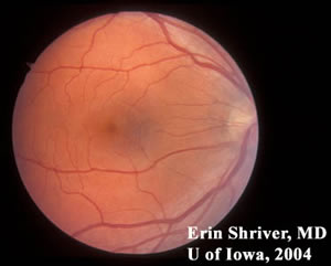
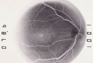
| Fundus Photo OD | Fundus Photo OS |
 |
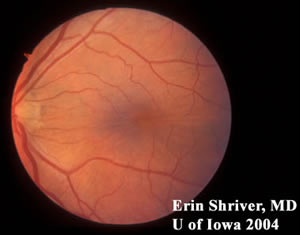 |
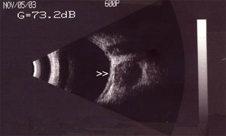
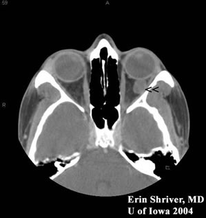
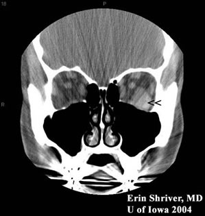
| Right eye had peripheral atrophic holes corresponding to the temporal scotoma on GVF. | Macular striae seen OS from intraconal mass. |
| OD (Normal) | OS |
 |
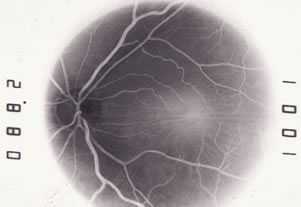 |
| Note: negative film FFA | Macular striae OS seen on FFA. |
 |
|---|
 |
 |
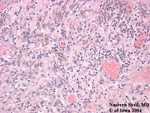
| Path Slide #1 | Path Slide #2 |
 |
 |
| Path Slide #3 |
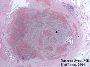
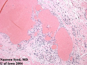
Path Slide #1: A large thrombus with inflammation (*) obscurred much of the large, dilated vascular spaces filled with blood. Immunohistochemistry confirmed that the thrombus contained mostly endothelial cells and fibroblasts, which ruled-out a neoplastic process. Thrombus formation is common because venous blood perfuses sluggishly through the cavernous hemangioma. Path Slide #2: Large, dilated vascular spaces filled with blood. Cavernous hemangiomas have pseudoencapsulated vascular channels separated by fibrous septa. Path Slide #3: Some inflammatory cells were found in this lesion. However, significant stromal inflammation and lymphoid follicles may support the diagnosis of a lymphangioma. |
EPIDEMIOLOGY
|
SIGNS
|
SYMPTOMS
|
TREATMENT
|
Shriver EM. Cavernous Hemangioma: 31-year-old caucasian female with a painless, dark spot in her left visual field. EyeRounds.org. Posted Feb. 21, 2005. Available from http://www.EyeRounds.org/cases/1-CavernousHemangioma.htm.

Ophthalmic Atlas Images by EyeRounds.org, The University of Iowa are licensed under a Creative Commons Attribution-NonCommercial-NoDerivs 3.0 Unported License.

Address
University of IowaLegal
Related Links