This site uses tracking information. Visit our privacy policy. Click to agree to this policy and not see this again.

Ophthalmology and Visual Sciences

Iris-Ciliary Body Melanoma:

57-year-old female with iris lesion

Iris-Ciliary Body Melanoma:

57-year-old female with iris lesion
Gina M. Rogers, MD, Nasreen A. Syed, MD, Wallace L. M. Alward, MD, Juan Fernandez de Castro, MD, Lauren Jensen, BS
January 15, 2010

Chief Complaint: Spot on the iris of the right eye.

History of Present Illness: A 57-year-old female noted a new tan-colored spot on the lower portion of her right iris while looking in the mirror. Her optometrist was concerned about this lesion, as it was easily visible and had not been noted on examinations over the prior three years. The patient denied any change in vision or other ocular symptoms.

Past Ocular History: Myopia and presbyopia

Medical History: Hypertension and depression

Ocular Exam:

  • Best corrected visual acuity: 20/20 OD and 20/20 OS
  • Pupils: Normal OU and symmetrically reactive, no RAPD
  • Applanation tonometry: 16 mm Hg OD, 15 mm Hg OS
  • Confrontation visual fields: full OU
  • Motility full and orthophoric
  • Slit Lamp Exam OD: See figures 1 and 2
  • Slit Lamp Exam OS was normal
Figure 1: Small lightly pigmented, slightly elevated lesion at the periphery of the iris of the right eye from approximately 6-8 o'clock. (click image for enlargement)
Figure 1.


Figure 2: Gonioscopy photos showing lesion extending to the periphery of the iris. There does not appear to be extension into the trabecular meshwork. (click image for enlargement)
Figure 2


Figure 3: Slit lamp and gonioscopy video demonstrates the lesions position in the periphery of the iris of the right eye with no apparent extension into the trabecular meshwork.

alternative video link: http://www.youtube.com/watch?v=AcYQZ-vW-jc


Figure 4: Echography of the right eye revealed a rounded lesion arising from the peripheral iris and extending below the iris plane. The lesion was 2.4mm thick and extended 3.9mm along the surface of the iris. It had low to medium reflectivity and appeared to be vascular. (click image for enlargement)
Figure 4

Based on the clinical appearance, it was thought that the patient's iris lesion was a melanoma.

The patient underwent a metastatic work-up for tumor staging. Liver function tests were at the upper limit of normal and a chest X-ray showed a nodule measuring less than 1cm in the lower lobe of the left lung. The patient subsequently underwent a chest CT to further evaluate the lung nodule, which was thought to be a granuloma. Given that she did not have any distant metastases, the decision was made to proceed with an iridocyclectomy.

Figure 5: Iridocyclectomy

alternative video link: http://www.youtube.com/watch?v=UTtGUKLNI6M

Also see Dr. Alward's Iridocyclectomy video at http://www.youtube.com/watch?v=QeG_mqfqKMA


Figure 6: Low power view of excised lesion demonstrating iris and ciliary body involvement. (click image for enlargement)
Figure 6


Figure 7: Higher power view of the lesion demonstrates infiltration of iris root and ciliary body with spindle cells and epithelioid cells, note none of the cells contain pigment. This is consistent with an amelanotic melanoma of the iris and ciliary body. Balloon cells with foamy cytoplasm are also present. (click image for enlargement)
Higher power view of the lesion demonstrates infiltration of iris root and ciliary body with spindle cells and epithelioid cells. Balloon cells with foamy cytoplasm are also present.
click on image for more detailed, unlabelled view


Figure 8: MART-1 (Melanoma Antigen Recognized by T-cells) stain: Positive brown staining of the lesion confirms the diagnosis of melanoma. Note the positive margins on the specimen. (click image for enlargement)
Figure 8

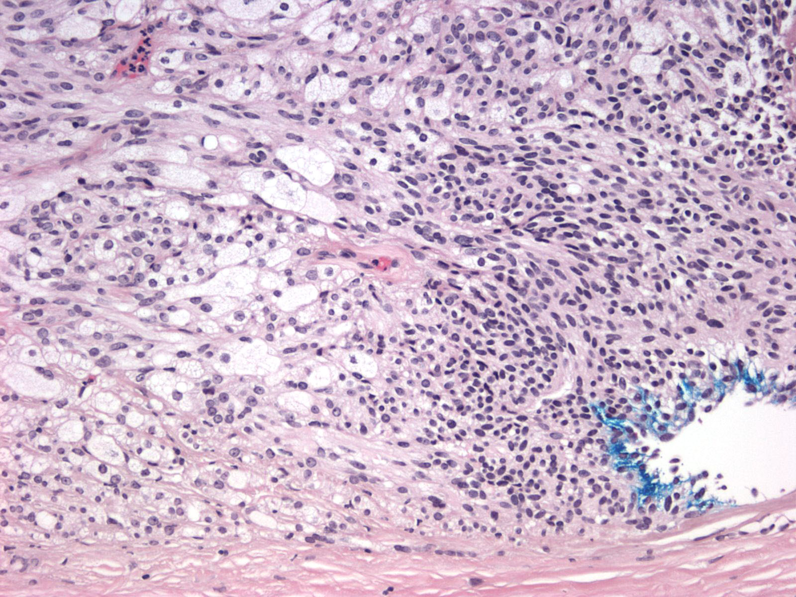
Based on the pathology, the lesion was an amelanotic melanoma involving the iris and ciliary body. It was not possible to determine if the tumor cells were ciliary body or iris in origin and it was concerning that the tumor margins were positive after what appeared grossly to be a complete resection. Given the rapid growth of the lesion and the involvement of the ciliary body (which carries a worse prognosis) it was thought to be in the best interest of the patient to treat the lesion aggressively. After discussion with the patient, the patient elected to have an enucleation of the affected eye. The patient underwent uncomplicated enucleation of the right eye in the following weeks.

She was evaluated by radiation oncology who determined that she did not have detectable distant metastases. They have recommended adjuvant radiation therapy to right eye socket, with repeat chest CT and liver function testing in 3-6 months. She has been tolerating radiation treatment well.

Figure 9: Pathology: Enucleated right globe s/p sector iridectomy. (click image for enlargement)
Figure 9

Discussion

Clinical diagnosis and treatment:

Uveal melanomas are the most common primary intraocular tumor in adults, most commonly occurring in the choroid. Iris melanomas represent 3-10% of uveal melanomas.(1) Clinically, small malignant melanomas of the iris can be very difficult to differentiate from benign iris nevi. They can range in appearance from off-white to dark brown. Usually iris melanomas are asymptomatic and are noticed by eye care specialists on routine examination. Of the uveal melanomas, the prognosis is best for iris melanomas as they have the lowest rates of metastasis and mortality.

There are several key clinical elements that may suggest malignancy on exam:

  • Dilated episcleral sentinel vessel
  • Growth of lesion anteriorly or erosion through iris root into anterior chamber
  • Growth into the lens which may produce sectoral or diffuse cataract, subluxation, lenticular astigmatism
  • Extension through sclera producing a dark epibulbar mass
  • Diffuse growth pattern extending around the circumference of the ciliary body, a ring melanoma.
  • Localized displacement of the iris anteriorly or infiltration of the trabecular meshwork which can produce tumor- induced glaucoma
  • Separation from and/or disruption of overlying ciliary epithelium, decreasing the production of aqueous humor and thus decreasing the intraocular pressure.

With respect to ancillary testing, echography is perhaps the most useful adjunct to the clinical exam. The A-scan technique allows the clinician to assess the lesion's internal reflectivity, vascularity and size. Melanomas typically have low internal reflectivity, a solid tumor pattern and can often have spontaneous vascular pulsations. The B-scan ultrasound can determine the size, position and thickness of the lesion in question.(2)

Given the relatively low rate of metastasis of an iris melanoma, management may involve close observation for signs of growth. Careful documentation and monitoring via photography and echography or ultrasound biomicroscopy is necessary.(3) Fine needle aspiration is sometimes used to obtain biopsy samples to make a tissue diagnosis. This method can yield both false positive and false negative results and does carry with it a small risk of tumor seeding. If it is decided that the lesion is quickly growing and suspicious, surgical resection is often accomplished by sector iridectomy or iridocyclectomy. Occasionally patients are treated with plaque radiotherapy although this method is usually reserved for patients who decline alternative interventions. The prognosis for iris melanoma is excellent as they usually do not metastasize. Lesions less than three clock hours in size have reported rates of extraocular spread of 3% at 5 years, 5% at 10 years, and 10% at 20 years with mortality rates estimated at 1-4%.(4,5)

Ciliary body melanomas grow behind the iris and thus may attain a large size before they are detected. Clinically they are visible only if the pupil is widely dilated or if they grow into the anterior segment. These lesions can remain asymptomatic until they grow enough to affect neighboring ocular structures; if extensive growth occurs patients may present with visual loss, photopsias, or visual field alterations.(6)

Ciliary body melanomas are associated with a worse prognosis than iris melanomas. These lesions have higher rates of metastasis with estimates of 25% at 5 years and 34% at 10 years.(7) The most common sites of metastases for both iris and ciliary body melanomas are the liver and lung. The initial metastatic workup involves checking liver function tests and obtaining a chest X ray. If any of these are abnormal, more extensive tests are undertaken, including liver ultrasound, CT-PET scan or MRI of the abdomen and chest. It has also been hypothesized that many have undetectable micrometastatic disease at the time of their primary diagnosis. For those with metastatic uveal melanoma, survival rates are poor, with a median survival of less than 6 months.(8) Because of the high rates metastases and poorer prognosis, ciliary body melanomas are treated more aggressively and patients often have to undergo enucleation for definitive therapy.

Histologic Diagnosis

The most important microscopic feature of uveal malignant melanomas that correlates with prognosis is the cell type. As per the Modified Callender Classification, lesions can contain predominantly spindle cells, epithelioid cells, or both.

  • Spindle Cells
    • A cells: (all A cells = nevus)
      • Spindle shaped cells with slender nuclei
      • Often with longitudinal nuclear fold
      • Fine chromatin and indistinct nucleous
    • B cells:
      • Spindle cells
      • Relatively plump, ovoid nuclei, prominent nucleolus
  • Epithelioid cells:
    • Large, more rounded or polyhedral cells with abundant cytoplasm
    • Lack of cohesion with adjacent cells.
    • Nuclei are large and round with prominent eosinophilic nucleoli
  • Mixed
    • Both spindle and epithelioid components

Several pathologic features correlate with prognosis: Spindle cell melanomas have the best prognosis, followed by mixed cell lesions. Epithelioid cell lesions generally have the worst prognosis. Size smaller than 10 mm in greatest dimension and the absence of extrascleral extension confer a better prognosis than larger lesions. Finally, the intrinsic microvascular pattern has implications for prognosis: A complex pattern associated with microvascular closed loops or networks (3 vascular loops located back-to-back) are associated with increased incidence of subsequent metastasis.(9, 10)

Diagnosis: Iridociliary Amelanotic Melanoma

EPIDEMIOLOGY

  • Malignant neoplasm arising from uveal tract melanocytes
  • Uveal melanomas are the most common primary intraocular malignancy in adults.
  • Overall incidence in U.S. is 6 cases/million
  • Average incidence by location(11)
    • 85% choroid
    • 10% ciliary body
    • 5% iris
  • Average age of onset: 40-50 years old(12)
  • Possible risk factors: Caucasian; light iris color; blond hair; ocular and oculodermal melanocytosis; and sunlight exposure(13, 14)

SIGNS

  • Spindle A cells: spindle-shaped cells with low nuclear-to-cytoplasmic ratio. No distinct nucleoli or mitoses. Lesions exclusively of spindle-A cell are considered nevi.
  • Spindle B cells: spindle-shaped cells with higher nuclear to cytoplasmic ratios and plump nuclei. Mitoses and nucleoli are present.
  • Epithelioid cells: large, more rounded or polyhedral cells with abundant cytoplasm, large nuclei, and prominent eosinophilic nucleoli; minimal cohesion with adjacent cells.
  • Mixed: combination of spindle and epithelioid cells.

SYMPTOMS

  • Choroid and ciliary body poorer prognosis than iris
  • Cell type: best to worst prognosis: Spindle > Mixed > Epithelioid
  • American Joint Committee on Cancer TNM Staging (www.mmmp.org) (15)

TREATMENT

  • Observation (iris only)
  • Surgical resection
  • Enucleation
  • Radioactive plaque
  • Charged-particle radiation

Differential Diagnoses, Iris Lesions:

  • Iris melanoma
  • Nevus
  • Melanocytoma
  • Metastasis to the iris
  • Iridic cysts
  • Iridocorneal endothelial syndrome
  • Melanocytosis: congenital ocular and oculodermal
  • Pigment epithelial hyperplasia or migration
  • Iridic component of underlying ciliary body melanoma
  • Peripheral anterior synechiae
  • Adenoma of the iris pigment epithelium
  • Medulloepithelioma
References
  1. Jakobiec FA, Silbert G. Are most iris melanomas really nevi A clinicopathologic study of 189 lesions. Arch Ophthalmol. 1981;99:(12)2117-2132.
  2. Marigo FA, Ginger PT, McCormick SA, et al. Iris and ciliary body melanomas: ultrasound biomicroscopy with histopathologic correlation. Arch Ophthalmol. 2000; 118(11):1515-1521.
  3. Alward, WLM. Glaucoma: The Requisites in Ophthalmology. Mosby; 2000,166-7.
  4. Augsburger, JJ, Correa, ZM, Freire, J, Brady, LW. Long-term survival in choroidal and ciliary body melanoma after enucleation versus plaque radiation therapy. Ophthalmology 1998; 105(9):1670-1678.
  5. Gragoudas, ES, Egan, KM, Seddon, JM, et al. Survival of patients with metastases from uveal melanoma. Ophthalmology 1991; 98(3):383-389.
  6. Albert DM, Miller JW, et al. Principles and Practice of Ophthalmology, Third Edition. Philadelphia: Saunders; 3609-3652, 4859-4874, 4875-4924.
  7. Shields JA, Shields CL. Intraocular Tumors: A Text and Atlas. Philadelphia: Sauders; 1992:137-153.
  8. Gass, JD. Observation of suspected choroidal and ciliary body melanomas for evidence of growth prior to enucleation. Ophthalmology 1980; 87(6):523-528.
  9. Folberg R, Mehaffey M, Gardner LM, Meyer M, Rummelt V, Pe'er J. The microcirculation of choroidal and ciliary body melanomas. Eye 1997; 11(2): 227-238.
  10. Pe'er J, Rummelt V, Mawn L, Hwang T, Woolson RF, Folberg R. Mean of the ten largest nucleoli, microcirculation architecture, and prognosis of ciliochoroidal melanomas. Ophthalmology 1994;101(7):1227-1235.
  11. COMS Collaborative Ocular Melanoma Study Group | (link to COMS web site) | (link to PubMed search)
  12. Gallagher, RP, Elwood, JM, Rootman, J, et al. Risk factors for ocular melanoma: Western Canada Melanoma Study. J Natl Cancer Inst 1985; 74(4):775-778.
  13. Albert, DM, Ryan, LM, Borden, EC. Metastatic ocular and cutaneous melanoma: a comparison of patient characteristics and prognosis. Arch Ophthalmol 1996; 114(1):107-108.
  14. Regan, S, Judge, HE, Gragoudas, ES, Egan, KM. Iris color as a prognostic factor in ocular melanoma. Arch Ophthalmol 1999; 117(6):811-814.
  15. Kujala, E, Kivela, T. Tumor, node, metastasis classification of malignant ciliary body and choroidal melanoma evaluation of the 6th edition and future directions. Ophthalmology 2005; 112(6):1135-1144.
Suggested citation format:

Rogers GM, Syed NA, Alward WLM, Fernandez de Castro J, Jensen L. Iris-Ciliary Body Melanoma: 57-year-old female with iris lesion. EyeRounds. org. January 15, 2010; Available from: http://www.EyeRounds.org/cases/104-Iris-Ciliary-Body-Melanoma.htm.


last updated: 01-15-2010

Image Permissions:
Creative Commons

Ophthalmic Atlas Images by EyeRounds.org, The University of Iowa are licensed under a Creative Commons Attribution-NonCommercial-NoDerivs 3.0 Unported License.