
Chief Complaint: 4 month-old female presents with an occlusive vascular lesion involving the right upper eyelid resulting in ptosis.
History of Present Illness: A strawberry-like, compressible area was first noted on the right upper eyelid within weeks of a normal delivery. The lesion has increased in size over the past three months. An intralesional injection of steroid was done without any resultant decrease in size of the mass. Topical and systemic steroid therapies were attempted therafter. On oral prednisone for almost a month, the child became increasingly irritable and there was no noted decrease in size of the lesion. Oral steroids were discontinued. The patient was continued on topical steroid cream only and referred to the University of Iowa Department of Ophthalmology for further evaluation and care. Throughout this time, the local ophthalmologist directed the parents to use part-time occlusion therapy of the child's left eye. At the time of referral, the parents were patching the left eye for 4 hours each day.
Past Ocular History: As noted above.
Medical History: Recent otitis media was treated with amoxicillin. The infant has two small hemangiomas on the toes.
Medications: Topical clobetasol cream (0.05%) applied generously to the right upper lid twice a day.
Family History: Noncontributory.
Social History: Noncontributory.
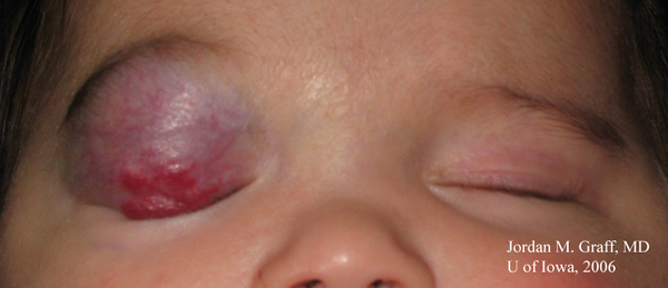 |
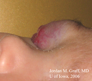
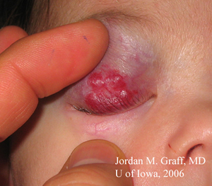
| A: Side view of the upper lid hemangioma demonstrates the elevated nature of the lesion. | B: Stretching the skin of the upper lid clearly shows two distinct portions of the lesion -- an elevated, red area where there is superficial hemangioma and a smooth, bluish-purple area representing a deeper component. |
 |
 |
Course: The examination and history are consistent with a capillary hemangioma of the right upper eyelid resulting in nearly total occlusion. Orbital echography demonstrated no intraorbital extension of the lesion. Due to the severity of the ptosis, amblyopia is a serious concern in this patient. Strict part-time occlusion of the left eye was continued and repeated intralesional steroid injections were offered. If the lesion continues to severely restrict vision in spite of repeated steroid injections, additional oral steroids or surgical excision should be considered.
Discussion: The history and presenting appearance of this patient are classic for capillary hemangioma. As the name implies, the lesions are made up of masses of very small-caliber vessels, and are created by abnormal growth of vascular endothelial cells. Therefore, capillary hemangiomas are hamartomas -- abnormal proliferations of tissue in a normal location. Unlike cavernous hemangiomas, which are usually found within the orbit, capillary hemangioma lesions tend to be less fibrotic, more cellular and typically unencapsulated. These lesions typically have a low rate of blood flow and are non-pulsatile.
In the vast majority of cases, the history and presentation of a periorbital capillary hemangioma is so typical as to be diagnostic. Caregivers will usually describe a growing red or purple spot that was not present at birth, but becomes larger and thicker over the first few months of life. The lesion will blanch with pressure, which is helpful in distinguishing it from the non-blanching port-wine stain of Sturge-Weber syndrome. While most hemangiomas are not present at birth, more than half of them will manifest by 1-2 months of age and 90-100% of the lesions will be present by 6-8 months of age. The peak size of the lesion is reached by 6-12 months, at which point the hemangioma - left untreated - will remain stable for a time before beginning to involute. In fact, spontaneous involution is the rule for untreated capillary hemangiomas, with progressive involution continuing until age 8. The classic teaching is that 40% of hemangiomas completely regress by age 4 and 80% completely regress by age 8. It is important to educate concerned parents about the natural course of involution. Nonetheless, a small proportion of these lesions can leave scars or other dermatologic changes to the tissues as they involute.
Treatment is indicated in cases of amblyopia, astigmatism or bony orbital asymmetry. Initiation of treatment for cosmetic deformity alone may be desired by the family, but this must be approached with caution given the natural course of involution and the potential risks of treatment.
Today, the first line treatment option is an oral beta blocker, such as propranolol. Topical beta blocker (timolol ophthalmic 0.5%) solution has also been used with good success. It is recommended that this be done in consultation with someone familiar with use of beta blockers in children. A preliminary heart rate, blood pressure and ECG are recommended before initiation of treatment. The most serious side effect to be monitored is hypoglycemia; other side effects include bronchospasm and bradycardia. In 2013 a consensus conference was held which offered suggestions on treatment of infantile hemangiomas (Drolet, 2013). In summation, initiation of treatment at a dose of 0.5-1.0 mg/kg/day of propranolol in twice (BID) to three times daily (TID) dosing is recommended. Then, after 1-2 weeks, this dose can be increased 0.5 mg/kg every 1-2 weeks targeted to a max dose of 2 mg/kg/day. Treatment is often continued for months until there is regression of the lesions.
In the past, first line therapy was intralesional steroid injection, with a mixture of long- and short-acting steroid (i.e., 40mg/ml of triamcinolone and 6mg/ml of betamethasone mixed in a syringe). The injection, if performed, should be through a medium-bore needle (i.e., 23-25 gauge) because smaller needles may clog with steroid particles. Though most hemangiomas will be reduced considerably with a single injection (see Figure 3), a repeat injection may be indicated at the 6th week of follow-up. Intralesional steroid treatment carries a risk of soft tissue atrophy, hypopigmentation, glaucoma, and possible systemic absorption. Additionally, there have been exceedingly rare reported cases of central retinal artery occlusion secondary to embolization of depot material from steroid injections (Egbert, 1996). A meta-analysis (Izadpanah, 2013) comparing corticosteroids versus propranolol showed a pooled response rate to treatment of 69 percent using corticosteroids versus 97 percent using propranolol .
An adjunctive therapy to intralesional steroids is oral steroids (prednisone 1-2mg/kg/day). Due to the increased risks associated with oral steroid therapy (e.g., agitation, changes in sleep pattern, increased risk of infection, osteoporosis, adrenal crisis, etc.), the assistance of a pediatrician, pediatric endocrinologist, or family physician is strongly encouraged. Oral steroids will likely be continued for months and must be tapered with caution due to the risk of adrenal crisis.
Surgical excision is an option as well. Surgery carries with it the risks of general anesthesia and recurrence of the lesion, but may help to significantly debulk large hemangiomas. Because of the complicated reconstruction that may be needed as well as risk of bleeding, referral to an oculoplastic surgeon is usually best. Throughout the treatment for vision-threatening complications of the hemangioma itself, the ophthalmologist needs to be concurrently minimizing the development of amblyopia. The consultation of a pediatric ophthalmologist may be helpful.
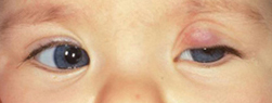
| A: Example of a similar patient with capillary hemangioma of the left upper lid. | B: Significant reduction in the lesion is seen after a single intralesional steroid injection. |
 |
 |
beam irradiation, interventional radiologic embolization, and various laser treatments (including CO2, argon, and Nd:YAG laser). Pulsed dye laser has also been used with some success, but only in very superficial lesions as usually this laser modality does not penetrate deeper than 1-2 mm. The use of topical steroid creams (e.g., clobetasol) has also been reported, but the response has been both slower and less impressive.
The ophthalmologist should be aware that many children with periorbital hemangioma may also have intraorbital extension of the lesion and/or systemic angiogenic lesions (including pulmonary, soft-tissue and/or skin lesions). B-scan ultrasonography of the orbit is helpful in determining the posterior extent of suspicious lesions while chest or abdominal imaging may uncover other hemangiomas. Some patients with large visceral capillary hemangiomas may develop thrombocytopenia, a condition known as Kasabach-Merritt syndrome. If the child bruises or bleeds easily or angiomatous lesions are suspected, a pediatrician should be involved in the work-up and treatment of the patient. Other conditions such as PHACE syndrome (PHACE = posterior fossa, hemangioma, arterial lesions, cardiac abnormalities, eye abnormalities - a cutaneous neurovascular syndrome characterized by large, segmental hemangiomas of the head and neck along with congenital anomalies of the brain, heart, eyes and/or chest wall) should also be considered.EPIDEMIOLOGY
|
SIGNSRed or purple elevated, subcutaneous soft mass that will blanch with pressure Hemangiomas are usually unilateral and located on the eyelid or brow
Reduced visual acuity may be noted (either due to amblyopia or uncorrected astigmatism from mass effect of a hemangioma on the cornea) May have both cutaneous and subcutaneous portions of the lesion
|
SYMPTOMSCare-givers usually notice a red or purple lesion
Amblyopia is seen in approximately 50% of patients with eyelid hemangioma
In rare cases with associated thrombocytopenia, a propensity for easy bruising or bleeding may be noted
|
TREATMENTSpontaneous involution is the rule - observation may be appropriate
In cases of occlusion amblyopia or significant astigmatism, prompt treatment should be initiated:
|
Lenci LT, Carter KD. Revision of [Graff JM, de la Garza AG, Carter KD. Capillary Hemangioma: 4 month-old female with occlusive vascular lesion of right upper eyelid. January 31, 2007.] EyeRounds.org. August 18, 2015; Available from: http://www.EyeRounds.org/cases/57-capillary-hemangioma-eyelid.htm.

Ophthalmic Atlas Images by EyeRounds.org, The University of Iowa are licensed under a Creative Commons Attribution-NonCommercial-NoDerivs 3.0 Unported License.

Address
University of IowaLegal
Related Links
