Chief Complaint: 30-year-old female referred for bilateral macular lesions.
History of Present Illness: 30-year-old female was found to have bilateral macular lesions on routine examination. She has no visual complaints. She denies any history of "night blindness", and her family history is negative for eye diseases.
PMH: healthy. FH: non-contributory.
EXAM
OD
|
OS
|
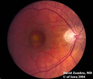
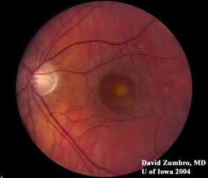
Best Vitelliform Macular Dystrophy
This is an autosomal dominant disease that results in bilateral vitelliform (egg-like) lesions in the macula. This disease is caused by mutations in the VMD2 gene that encodes a chloride channel in the basolateral membrane of the retinal pigment epithelium (RPE), resulting in lipofuscin deposits in the RPE layer. The defective chloride channels result in an abnormal electrooculogram (EOG), i.e. low Arden ratio = light peak/dark trough.Patients will have bilateral, symmetric vitelliform lesions. Visual acuities are usually remarkably better than expected for patients with large macular lesions. A pseudohypopyon (aqueous-lipid fluid level) can be seen in the above lesions.
Best lesions may be associated with choroidal neovascular membranes and may have sub-retinal hemorrhage. Hemorrhage may also occur after minor globe trauma. The natural history is full recovery of vision after hemorrhage; thus, sub-foveal surgery is not indicated because patients who have undergone surgery do much worse than patients who are allowed to heal without surgical intervention.Visual potential is good. According to Dr. Edwin Stone, 95% of Best Dystrophy patients will have vitelliform lesions by age 40, and 75% of Best Dystrophy patients will have driving vision at age 60. Without a family history, only 25% of patients will have a mutation. The other patients will have another retinal dystrophy, e.g., pattern dystrophy (RDS gene), cuticular drusen, pigment epithelial detachment, central serous retinopathy, etc..
EPIDEMIOLOGY
| SIGNS
|
SYMPTOMS
| TREATMENT
|
Doan A, Stone EM: Best Vitelliform Macular Dystrophy: 30-year-old female referred for bilateral macular lesions. February 21, 2005; Available from: http://www.EyeRounds.org/cases/case11.htm.

Ophthalmic Atlas Images by EyeRounds.org, The University of Iowa are licensed under a Creative Commons Attribution-NonCommercial-NoDerivs 3.0 Unported License.

Address
University of IowaLegal
Related Links