Choroidal Osteoma:
10-year-old girl presents for annual follow-up. She reports that she recently failed a school screening
Choroidal Osteoma:
10-year-old girl presents for annual follow-up. She reports that she recently failed a school screening
Chief Complaint: 10-year-old girl presents for annual follow-up. She reports that she recently failed a school screening.
History of Present Illness: However, the patient offers no complaints herself.
PMH: healthy. FH: non-contributory.POH: Limbal Dermoid OS, Retinal Lesion OD.
EXAM
- Best corrected visual acuities: 20/40 OD, 20/20 OS
- Stereo Vision: normal
- Pupils: normal, no RAPD
- VF: full to CF OU
- EOM: normal
- IOP: normal OU
- SLE: normal
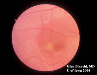
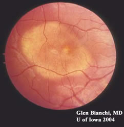
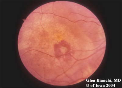
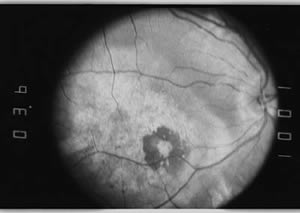
- DFE: see below
Discussion
Choroidal Osteomas
This is an osseous choristoma of the choroid. There is no known treatment. There is a predilection for women, and it is typically found in the second or third decade of life. Choroidal osteomas may be bilateral in 25% of cases. Growth is seen in 40% of cases with long-term follow-up. These lesions may be associated with choroidal neovascular membranes (CNVM), sub-retinal fluid, and sub-retinal hemorrhages.
80% of patients have 20/30 vision or better at presentation. Visual loss may be gradual or acute due to RPE degeneration or CNVM. Dr. Gass estimates that up to 33% of eyes with choroidal osteoma may develop CNVM.
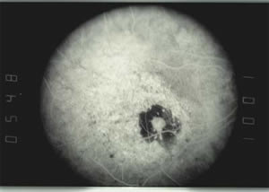
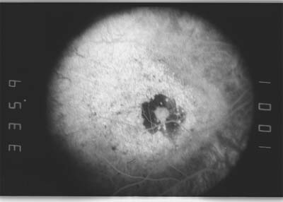
Fluorescein Angiography: Early, patchy hyperfluorescence with intense late staining.
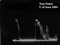
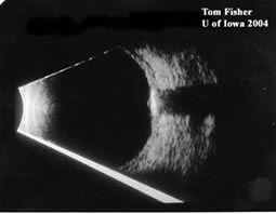
Echography : Densely echogenic (extremely high internal reflectivity of bone), persists at low sensitivities. Marked "Shadowing," or sound attenuation posterior to lesion. "Void" appears behind lesion.
Histology : Mature bone with interconnecting marrow spaces. Lesion is sharply demarcated form choroid.
Diagnosis: Choroidal Osteomas
EPIDEMIOLOGY
- Rarely familial.
- Predilection for women.
- Presents in second and third decade of life.
- Incidence: rare
|
SIGNS
- Bilateral in 25% of cases.
- About one-third of cases will be associated with a CNVM.
- Yellow-white lesion, well demarcated.
- Fluorescein Angiography: Early, patchy hyperfluorescence with intense late staining.
- Echography : Densely echogenic (extremely high internal reflectivity of bone), persists at low sensitivities. Marked "Shadowing," or sound attenuation posterior to lesion. "Void" appears behind lesion.
|
SYMPTOMS
- Mild vision loss in the early stages.
- Moderate to severe vision loss if associated with RPE atrophy or CNVM.
|
TREATMENT
- No treatment available for choroidal osteoma.
|
Differential Diagnoses
- Amelanotic Choroidal Melanoma
- Amelanotic Choroidal Nevus
- Choroidal Hemangioma
- Metastatic Carcinoma
- Organized Submacular Hemorrhage
- Cicatricial Macular Degeneration
References
- Aylward, G.W., et al., A long-term follow-up of choroidal osteoma. Arch Ophthalmol, 1998. 116(10):1337-41.
- Shields, C.L., J.A. Shields, and J.J. Augsburger, Choroidal osteoma. Surv Ophthalmol, 1988. 33(1):17-27.
- Grand, M.G., et al., Choroidal osteoma. Treatment of associated subretinal neovascular membranes. Retina, 1984. 4(2):84-9.
- Gass, J.D., et al., Choroidal Osteoma. Arch Ophthalmol, 1978. 96(3):428-35.
- Kida, Y., et al., Choroidal osteoma in an infant. Am J Ophthalmol, 1997. 124(1):119-20.
- Fava, G.E., et al., Choroidal osteoma in a 6-year-old child. J Pediatr Ophthalmol Strabismus, 1980. 17(3):203-5.
- Mizota, A., R. Tanabe, and E. Adachi-Usami, Rapid enlargement of choroidal osteoma in a 3-year-old girl. Arch Ophthalmol, 1998. 116(8):1128-9.
- Katz, R.S. and J.D. Gass, Multiple choroidal osteomas developing in association with recurrent orbital inflammatory pseudotumor. Arch Ophthalmol, 1983. 101(11):1724-7.
- Okada, K., et al., Bilateral choroidal osteomas associated with histiocytosis X. Jpn J Ophthalmol, 1996. 40(1):111-5.
- Trimble, S.N. and H. Schatz, Choroidal osteoma after intraocular inflammation. Am J Ophthalmol, 1983. 96(6):759-64.
- Eting, E. and H. Savir, An atypical fulminant course of choroidal osteoma in two siblings. Am J Ophthalmol, 1992. 113(1):52-5.
- Noble, K.G., Bilateral choroidal osteoma in three siblings. Am J Ophthalmol, 1990. 109(6):656-60.
- Teich, S.A. and J.B. Walsh, Choroidal osteoma. Ophthalmology, 1981. 88(7): p. 696-8.
- Gass, J.D., New observations concerning choroidal osteomas. Int Ophthalmol, 1979. 1(2): p. 71-84.
- Avila, M.P., et al., Bilateral choroidal osteoma with subretinal neovascularization. Ann Ophthalmol, 1984. 16(4):381-5.
- Augsburger, J.J., J.A. Shields, and C.J. Rife, Bilateral choroidal osteoma after nine years. Can J Ophthalmol, 1979. 14(4):281-4.
- Morrison, D.L., et al., Review of choroidal osteoma: successful krypton red laser photocoagulation of an associated subretinal neovascular membrane involving the fovea. Ophthalmic Surg, 1987. 18(4): p. 299-303.
- Rose, S.J., J.F. Burke, and R.J. Brockhurst, Argon laser photoablation of a choroidal osteoma. Retina, 1991. 11(2):224-8.
- Burke, J.F., Jr. and R.J. Brockhurst, Argon laser photocoagulation of subretinal neovascular membrane associated with osteoma of the choroid. Retina, 1983. 3(4):304-7.
- Shields, J.A., et al., Ocular manifestations of the organoid nevus syndrome. Ophthalmology, 1997. 104(3): p. 549-57.
- Traboulsi, E.I., et al., Posterior scleral choristoma in the organoid nevus syndrome (linear nevus sebaceus of Jadassohn). Ophthalmology, 1999. 106(11):2126-30.
Suggested citation format:
Bianchi GM, Keech RV, Olson R: Choroidal Osteoma: 10-year-old girl presents for annual follow-up. She reports that she recently failed a school screening. February 21, 2005; Available from: http://www.EyeRounds.org/cases/case12.htm.
last updated: 02-21-2005
Image Permissions:

Ophthalmic Atlas Images by EyeRounds.org, The University of Iowa are licensed under a Creative Commons Attribution-NonCommercial-NoDerivs 3.0 Unported License.