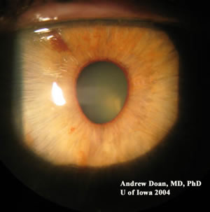
Chief Complaint: 63-year-old female with PAS, "iris mass", corectopia, and increased IOP OS.
History of Present Illness: Previously healthy female with progressive corectopia and PAS formation OS over the last year. IOP in mid-20s OS. Patient consulted for evaluation of "iris mass".
PMH/FH/POH: non-contributory.
| OS | Gonioscopy OS |
|
|
|
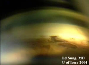
| Corectopic (i.e., oval) left pupil . | Broad area of peripheral anterior synechiae (PAS) at 6:00 o'clock position with area of iris atrophy and pigmented iris stroma. |
| Cornea OS | Cornea OS |
|
|
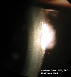
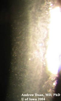
| Cornea with hammered silver appearance of endothelium. | Magnified view: cornea with hammered silver appearance of endothelium. |
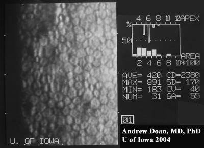
Endothelium OD |
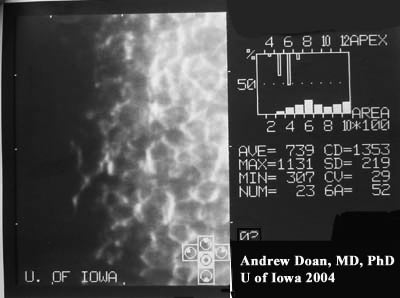
Endothelium OS |
|
|
| Normal, hexagonal shaped cells. Normal cell count (2380) | Abnormal endothelium with large spaces between cells and low cell count (1353). The color of the cells are also reversed with black surrounded by a white border. |
This patient presented with unilateral corectopia, PAS, hammered silver appearance of the corneal endothelium OS, areas of iris atrophy OS, and history of intraocular pressure spikes OS. The unilaterality of the presentation must include a differential diagnosis of unilateral glaucomas, which can be divided into glaucomas with heterochromia and those without heterochromia.
Specular microscopy demonstrated an abnormal corneal endothelium with low cell counts OS. This case is an example of early essential iris atrophy. This patient has an increased risk of developing glaucoma OS and must be routinely followed with intraocular measurements, optic nerve exam, gonioscopy, and visual field testing.
ICE syndrome is a spectrum of diseases. There are three clinical categories: Chandler's syndrome, Cogan-Reese syndrome, and essential iris atrophy. The unifying abnormality in all three entities is the abnormal, unilateral, hammered silver appearance of the corneal endothelium. Chandler's syndrome presents with only the hammered silver appearance of the corneal endothelium. Cogan-Reese syndrome presents with the corneal findings and pigmented nodules on the iris. Essential iris atrophy presents with the corneal findings and corectopia with developments of stretch and melt holes with disease progression.
EPIDEMIOLOGY
|
SIGNS
|
SYMPTOMS
|
TREATMENT
|
Doan A, Alward W: Iridocorneal Endothelial Syndrome (ICE) - essential iris atrophy : 63-year-old female with PAS, "iris mass", corectopia, and increased IOP OS. February 21, 2005; Available from: http://www.EyeRounds.org/cases/case14.htm.

Ophthalmic Atlas Images by EyeRounds.org, The University of Iowa are licensed under a Creative Commons Attribution-NonCommercial-NoDerivs 3.0 Unported License.

Address
University of IowaLegal
Related Links