35-year-old female with blurry vision OU lasting hours in the morning
Fuchs Endothelial Dystrophy:
35-year-old female with blurry vision OU lasting hours in the morning
Andrew Doan, MD, PhD, Andrew Lee, MD
February 21, 2005
Chief Complaint: 35-year-old female with complaint of intermittent blurry vision OU lasting hours.
History of Present Illness: Patient was sent to neuro-ophthalmology for evaluation of blurry vision OU. The patient complained of decreased vision upon awakening that is not associated with pain. She stated that it's difficult for her to read. The patient also mentioned that her vision seems to clear up as the day progresses.
EXAM:
Vision: 20/25 OU at distance and near.
Pupils: 5 mm in dark, 2 mm in light, no RAPD.
Extraocular Motility: Full motility without pain.
IOP: 18 mmHg OU.
Visual Field: Full OU.
DFE: normal macula, vessels, and periphery OU.
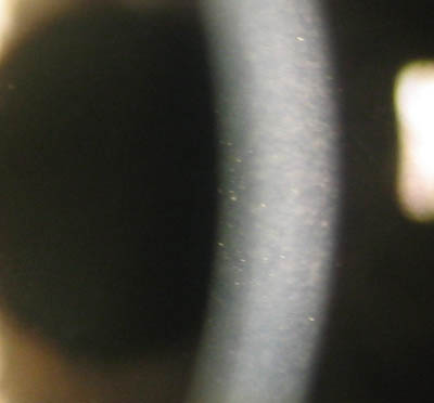
SLE: notable for a beaten metal appearance of the corneal endothelium OU
Slit beam through cornea. Notice the imperfections (guttae) of the corneal endothelium on the left side of the beam.
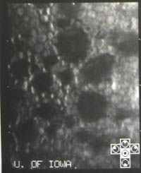
Specular microscopy showed low endothelial cell count. Less than 1800 cells/mm2. Most people have more than 2000-3000 cells/mm2 (average of 2400 cells/mm2)
The diagnosis is Fuchs endothelial dystrophy due to endothelial cell loss. Patients have worse vision in the morning because of the eyes being closed during the night causing a buildup of corneal edema (because the endothelium helps keep the cornea dry and clear). During the day, with the eyes open, the cornea becomes a little more dehydrated. Patients can be treated with dehydrating ointments (5% NaCl) or even with a hair dryer in the earlier symptomatic stages prior to PK.
The dark areas on the specular microscopy are regions where the endothelial cells have died (guttae). Because these cells are terminally differentiated cells, they do not divide, so the remaining cells have to stretch to cover more area.
Diagnosis: Fuchs endothelial dystrophy
EPIDEMIOLOGY
Incidence unknown.
May have family history.
Females:Male ratio is 3:1
Age of onset: over age 50.
Mild vision problems may develop in younger patients with central cornea guttae.
SIGNS
Cornea guttae (beaten metal appearance)
Cornea stromal edema
Bilateral with possible asymmetry
Cornea bullae.
Folds in Descemet's membrane.
Cornea scarring in the late stages.
Low endothelial cell counts on specular microscopy.
SYMPTOMS
Glare and blurred vision, typically worse in the morning due to corneal edema from lids being closed at night.
Pain with severe corneal decompensation.
TREATMENT
Topical NaCl 5% drops 4X/day and ointment at night to help dehydrate the cornea.
Reduce IOP with anti-glaucoma medications to reduce cornea edema.
Topical steroids may help.
Ruptured bullae should be treated as a corneal abrasion.
Mild disease may be treated with gentle blow drying of cornea in the morning.
Caution with intraocular surgeries that may injure the corneal endothelium, e.g. cataract surgery. BSS(+) [with glutathione] may help reduce loss of endothelial cells during cataract surgery.
Differential Diagnoses
Pseudophakic bullous keratopathy
Chandler's syndrome (this is a unilateral ICE syndrome with a hammered silver appearance of the corneal endothelium)
Posterior polymorphous dystrophy
Congenital hereditary endothelial dystrophy (present at birth)
Doan A, Lee AG: Fuchs Endothelial Dystrophy: 35-year-old female with blurry vision OU lasting hours in the morning. February 21, 2005; Available from: http://www.EyeRounds.org/cases/case5.htm.
University of Iowa
Roy J. and Lucille A. Carver College of Medicine
Department of Ophthalmology and Visual Sciences
200 Hawkins Drive
Iowa City, IA 52242
University of Iowa
Roy J. and Lucille A. Carver College of Medicine
Department of Ophthalmology and Visual Sciences
200 Hawkins Drive
Iowa City, IA 52242