This site uses tracking information. Visit our privacy policy. Click to agree to this policy and not see this again.

Ophthalmology and Visual Sciences

Assessment and Management of Ocular Trauma

Assessment and Management of Ocular Trauma

Written by: Sudeep Pramanik, M.B.A., M.D.
With contributions by: Andrew Doan, M.D., Ph.D and
Thomas A. Oetting, M.D.,

The University of Iowa
Department of Neurology and Department of Ophthalmology and Visual Sciences


INITIAL EXAM OF OCULAR AND ADNEXAL INJURIES

OPEN GLOBES

  • If you suspect an open globe, you may perform a quick exam before taking the full history.
  • If the globe is ruptured, call the senior resident and finish the history/paperwork while backup is on its way.
  • Make sure the patient has updated tetanus immunization, order IV antibiotics/narcotics/antiemetics, call OR/anesthesia, ask about time of last meal, and prepare admission paperwork.
  • Place shield over eye when not examining. Do NOT patch the eye; instruct transferring caregiver NOT to patch the eye.
  • Inspect eyes without placing pressure on the globe (e.g., use Desmarres retractors).
  • Signs of open globe include:
    1. penetrating lid injury,
    2. bullous subconjunctival hemorrhage
    3. shallow anterior chamber
    4. blood in the anterior chamber (hyphema),
    5. peaked pupil
    6. iris disinsertion (iridodialysis)
    7. lens dislocation, and
    8. vitreous hemorrhage. Loss of red reflex can indicate vitreous hemorrhage or retinal detachment.
  • Palpate for discontinuity of the orbital rim and detachment of the medial canthal tendon, signs of severe orbital trauma.
  • A positive Seidel test will help detect leaking aqueous or exposed vitreous. Fluorescein ophthalmic strips are wetted with normal saline. Concentrated fluorescein is dark orange, but if it becomes diluted with aqueous, it turns bright green under blue light.
  • The presence of an intraocular foreign body (IOFB) suggests globe penetration. Plain film x-rays of the head and/or CT scan will help determine the location and presence of a metallic IOFB. Do NOT do a MRI scan if an IOFB is suspected. Additionally, a CT scan will help show the shape and integrity of the traumatized globe.
  • If globe is obviously open, do not check pressure, motility, nor dilate the traumatized eye.
  • Avoid ointment on an open globe. Non-preserved antibiotics (e.g. moxifloxacin) are okay if there is a delay in getting to the OR.
  • Advise referring clinicians to place a shield over the injured eye (no patch), administer anti-nausea and pain medicine (if caregiver is a physician), and send the patient. No need to delay transfer for IV antibiotics.

Clinical Signs of Severe Globe Trauma and Open Globe

Bullous Subconjunctival Hemorrhage
Bullous subconjunctival hemorrhage in this patient resulted from a fall and may signify severe orbital trauma resulting in globe rupture.
Bullous Subconjunctival Hemorrhage
Bullous subconjunctival hemorrhage in this patient resulted from a fight and resulted in severe globe rupture extending posteriorly and could not be repaired. The eye was enucleated within two weeks to reduce the risk of sympathetic ophthalmia.
Hyphema
Hyphema
The presence of red blood cells in the AC may layer out into a hyphema (white arrow head).
Peaked Pupil
Peaked Pupil
A penetrating wound from a knife resulted in a peaked pupil (corectopia) pointing towards the corneal-scleral defect. There was iris in the corneal-scleral wound. The open globe was repaired with 10-O nylon, and the patient did well subsequently.
Iridodialysis
Iridodialysis
The tissues of the eye are organized into concentric rings like an inner tube of a tire. When there is blunt trauma, increased intraocular pressure forces the globe to expand which places stress on the concentric rings causing them to stretch. With sufficient force, the tissues can tear. The black arrow denotes an inferior iris tear from blunt trauma (iridodialysis).
Retinal Detachment
Retinal Detachment
Severe ocular trauma may result in retinal detachments. This patient had a penetrating globe injury while mowing the lawn without safety glasses. The detached retina can be seen as the yellowish-orange mass behind the iris.
Deflated Globe
Deflated Globe
Deformed eye due to abrupt globe rupture after walking into a door. The patient had a corneal graft in the left eye that ruptured due to blunt trauma.
Seidel Test
Seidel Test
Concentrated fluorescein is dark orange but turns bright green under blue light after dilution. This patient had a penetrating eye injury with vitreous loss. The site was Seidel positive with streaming fluorescein, and the protuding vitreous stained green too.
Intraocular Foreign Body (IOFB)
This patient had a penetrating eye injury from a metal fragment while hammering metal on metal without safety glasses. Retroillumination (i.e. projection of light into the pupil) detects the red glow of the retina via retroillumination through a superior-nasal iris defect (white arrow). He had a self-sealed corneal wound that was NOT Seidel positive.
Plain film X-Ray of IOFB
X-Ray of the head shows a bright, well-defined, round signal in the right orbit. This is the metallic IOFB in the patient shown on the left. The patient had a pars plana vitrectomy and removal of the IOFB by the retina service.
Shotgun Injury to the Head
Shotgun injury to the right eye with hyphema (iris details are obscurred), soft globe, and bullous subconjunctival hemorrhage. The eye was ruptured beyond repair and was enucleated.
CT Scan of Shotgun Injury
Axial CT scan of the head denoted several subcutaneous and intracranial shotgun pellets (black arrows). The right globe is grossly deformed without a lens and continuous scleral signal (compared to left eye).
Ruptured Globe from Hit & Run
This patient had a globe rupture after being hit by a car. The right globe is proptotic and soft. The AC was filled with blood which made the cornea appear black with obscuration of iris details.
Axial CT Scan of Hit & Run Case
The blue arrow denotes a severely deformed right eye with hyper-dense signals within the globe (vitreous hemorrhage and blood clot).

FOREIGN BODIES

  • Remove non-impaled conjunctival foreign bodies (FB) using irrigation ± wet cotton swabs.
  • Remove corneal FB with wet cotton swabs if you are sure it does not penetrate the cornea.
  • Quinolone four times daily ± bandage contact lens.
  • Follow next day.
  • If infiltrate develops, then video/take photographs, culture, perform confocal microscopy if available, and call the senior resident.

CORNEAL ABRASIONS

  • Evert and sweep the lids/irrigate for residual foreign body.
  • Debride loose epithelium with jewelers forceps.
  • Scopolamine 0.25% twice daily if iritis is present.
  • Acular® (ketorolac tromethamine) four times daily for 3 days can help with pain control.
  • Quinolone four times daily and bandage contact lens may help with healing and comfort.
  • Follow next day.
  • If infiltrate develops, then record video/take photographs, culture, perform confocal microscopy if available, and call the senior resident.
  • Use topical anesthesia for exam only. Make sure the patient does not leave with anesthetic drops, these cause serious keratopathy with chronic use.
  • Give narcotics to patients in severe pain.
  • ALWAYS DILATE AND EXAMINE THE NON-TRAUMATIZED EYE.

CHEMICAL BURNS

  • For chemical burns, injury grade is based on exam before irrigation has begun (it is rare that you will see patients before irrigation is initiated).
  • Start irrigation IMMEDIATELY with Normal Saline or Lactated Ringers Solution (2 liters)—wait 5 minutes and check pH.
  • Additional irrigation as needed until pH is 7.0-7.5.
  • Continue irrigation until pH is normal—compare pH to that of the other eye because often pH paper will read either too alkaline or acidic.
  • Sweep for particulate matter and examine fornices.
  • Check intraocular pressure (IOP).
  • Call the senior resident.
  • Note limbal ischemia, epithelial defect, stromal loss/haze, edema, anterior chamber depth, inflammation, and lens clouding.

RETROBULBAR HEMORRHAGE

  • If patient has retrobulbar hemorrhage with decreased acuity, increased IOP, check central retinal artery perfusion; you may need to perform a lateral canthotomy/cantholysis.
  • Call the senior resident.
  • If you have doubt about a relative afferent pupillary defect (RAPD) and need a better look at the posterior segment, dilate one eye. A RAPD can be checked by reverse pupil exam.

TRAUMATIC OPTIC NEUROPATHY

  • For traumatic optic neuropathy, the usual mechanism is a blow to the superior rim with force transmitted to the optic canal.
  • CT without contrast to look for bone fragment on optic nerve or sheath hematoma (emergent surgical intervention necessary).
  • IV steroids effectiveness is unproven, but may be given in the absence of contraindications.
  • Based on spinal cord injury studies, methylprednisolone IV can be given: 30 mg/kg loading dose then 5.4 mg/kg/hr for 24 hours (if started within 3 hours of injury) or for 48 hours (if started 3-8 hours after injury).
  • There is no consensus for the steroid dosage and treatment regimen in traumatic optic neuropathy.

WHITE-EYED & OTHER BLOWOUT ORBITAL FRACTURES

  • White-eyed blowout fracture in children with motility restriction from entrapment of tissues can lead to ischemia and fibrosis, so it should be repaired ASAP (<48 hrs).
  • Other types of orbital fracture repair can wait 5-7 days for soft-tissue swelling to decrease.
  • Floor and medial wall fractures are repaired if there is significant enophthalmos, (sunken eyes >2mm), large fracture (>50% of the orbital floor), entrapment on CT, or restriction (positive forced ductions or oculocardiac reflex).
  • Have patient avoid nose blowing or sneezing (applying pressure to upper lip may reduce sneezing).
  • Call your senior resident.
  • Record patient information and contact number and KEEP THE FILMS.
  • Tell the oculoplastics fellow or staff the next morning and give them the films.
  • Le Fort I fractures do not involve the orbit, Le Fort II and III involve nasolacrimal system. Check for facial sensation (Cranial Nerve V1 = orbit roof and V2 =orbit floor) and trismus (pain and tonic contraction of the muscles of mastication). If trismus is present, then suspect a zygomatic maxillary complex (ZMC) fracture.
  • Nasal-orbital-ethmoid (NOE) fractures can cause traumatic optic neuropathy and involve the nasolacrimal system.

LID LACERATIONS

  • With lid lacerations, look carefully for fat prolapse, lid margin involvement and canalicular involvement.
  • For lid laceration repair, get General Ophthalmic Surgery tray and suture suitcase from minor room. 7-O Vicryl is used for skin and 5-O Vicryl is used for deep closure.

ORDERING IMAGING STUDIES

  • In the ER, order CT scan of the orbits with fine cuts (1-2mm) and true coronals, this is particularly important for IOFB.
  • Coronal reconstructions and maxillo-facial CT scans are often adequate if already done from a transferring ER.
  • Avoid MRI scans if IOFB or intracranial FB suspected.

Examples of Various Orbital Fractures

Nasal-orbital-ethmoid (NOE) Fracture
The patient suffered a softball injury to the forehead with subsequent fracture and compression of his ethmoid sinus, i.e., nasal-orbital-ethmoid (NOE) fracture. Notice the bridge of the nose is pushed-in and compressed. The nose and ethmoid sinus provides a "speed bumper" for the brain. The compression of the nasal-orbital-ethmoid complex reduces injury to the delicate intracranial structures.
Orbital Floor and Medial Wall Fracture
Coronal CT Scan of the Face & Orbits
The patient had a floor fracture (white arrowhead) and blood in the left maxillary sinus. The medial wall of the left orbit is also fractured.
Zygomatic maxillary complex (ZMC) Fracture
Coronal CT Scan of the Face & Orbits
The patient had a zygomatic maxillary complex (ZMC) fracture. The arrowheads denote fractures along the zygoma, orbital floor, and maxilla. The left maxillary sinus is filled with blood.
Orbital Roof Fracture
Coronal CT Scan of the Face & Orbits
The patient suffered a large orbital roof fracture (white arrow) associated with marked intracranial hemorrhage.
Medial Wall Blowout Fracture
Axial CT Scan of the Orbits
The patient was in an altercation and developed a medial wall blowout fracture (white arrowhead) with entrapment of the medial rectus muscle. The medial wall fracture was surgically repaired. Compare the medial wall integrity of the left orbit with the right orbit.

WHAT SUTURES DO I NEED?

Repair Suture
Cornea 10-0 Nylon on spatulated needle
Sclera 9-0 or 8-0 Nylon on spatulated needle
Conjunctiva 7-0 Vicryl
Medial canthal tendon 4-0 Mersiline
Skin 5-0 Fast Absorbing Gut (preferred at Iowa), 7-0 Vicryl or 6-0 Silk
Lid margin

4-0 Silk through tarsal plate + 6-0 Silk for skin

-OR-

5-0 Vicryl through tarsal plate and two 7-0 Vicryl sutures for lid margin (vertical matress) and skin

Canalicular laceration

Stent with silicone tubing (leave for 6 months)

6-0 Chromic and 6-0 Silk sutures

SUTURE PLACEMENT PRINCIPLES

  • Entry and exit points - equidistant from the wound edge.
  • Entry and exit - vertical to the surface.
  • Sutures - perpendicular to the wound.
  • Sutures - passed through 90% corneal depth and buried in the cornea.
  • Cornea - closed from the periphery to the center.
  • Sutures - longer near the limbus and shorter toward the central cornea.
  • Shorter sutures - spaced closer together.
  • Align anatomic landmarks first (e.g., limbus).
  • Avoid placing sutures in the visual axis if possible.
  • Scleral sutures - 75-90% deep, with entry and exit at least 1 mm from the wound edge.
  • Start anterior and progress posterior, so the wound is more stable when you need to rotate the globe for posterior access.
  • Attempt to close the entire wound, unless accessing its posterior extent would result in loss of additional ocular contents.
  • Rotate scleral knots posteriorly and bury conjunctival sutures if possible

EXAMINING FACIAL FRACTURES

Type of Fracture… Look for…
Le Fort II-III Optic canal and nasal lacrimal duct injury
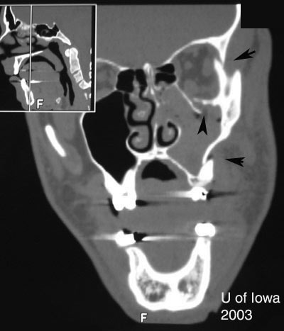
Orbital floor fracture Vertical diplopia, extraocular muscle rounding on CT, V2 hypoesthesia, orbital/sub-cutaneous emphysema, enophthalmos, oculocardiac reflex, pupil abnormalities
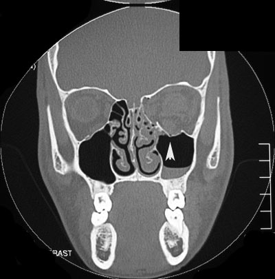
Medial wall fracture (orbit) Horizontal diplopia, orbital emphysema, orbital hemorrhage, enophthalmos
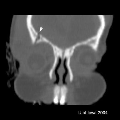
Roof fracture (orbit) Restricted upgaze, ptosis, epistaxis, CSF rhinorrhea, anosmia, depression of superior rim, V1 hypoesthesia, hypo-ophthalmos, pulsatile exophthalmos; traumatic optic neuropathy
ZMC Fracture Point tenderness, ecchymosis, malar flattening, lateral canthal downward displacement, V2 hypoesthesia, trismus, malocclusion of jaw, inferior or lateral rim step-off.
NOE (Nasal-Orbital-Ethmoid) Fracture Facial flattening, telecanthus, epistaxis, CSF rhinorrhea, anosmia, nasolacrimal duct damage, optic canal damage

ADMINISTERING LOCAL NERVE BLOCKS

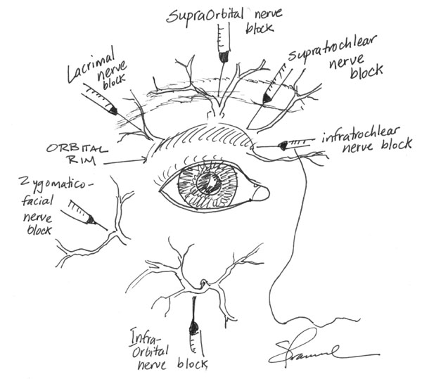
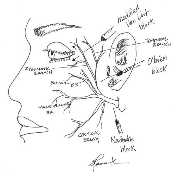
The most common local anesthetic mixture is 2% lidocaine with 1:100,000 epinephrine to provide some hemostasis. Addition of 0.5% bupivacaine will provide longer anesthesia for lengthy procedures. The following diagrams illustrate common local nerve blocks used in ophthalmology.

Common Periorbital Nerve Blocks
Common Facial Nerve Blocks

ADMINISTERING RETROBULBAR BLOCK

Retrobulbar blocks are useful methods of achieving anesthesia for intraocular and orbital surgeries. Blocks are good alternatives to general anesthesia when general anesthesia is undesireable or contraindicated.

There are many techniques to adminster a retrobulbar block. The method described here is what I prefer. Depending on the type of anesthetic, a block may last over four hours with a mixture of lidocaine 1% and bupivacaine 0.375%.

  • Clean the lower lid with an alcohol wipe.
  • Have the patient look straight ahead.
  • Use a 5 cc syringe and a 23 gauge, 1.5 inch flat grind needle.
  • Start 2/3 lateral along the inferior orbital rim (inferior to the temporal limbus) with the needle tip pointing perpendicular to the plane of the patient’s face. Place the index finger between the globe and the infraorbital rim, elevating the globe.
  • Stabilize with the little finger and hypothenar eminence firmly on the bones of the patient’s cheek.
  • Enter just inferior to the globe and perpendicular to the plane of the face.
  • Once you feel the first pop through the orbital septum, angle 45 degrees medially and 45 degrees superiorly towards the apex of the orbit until the second pop throught the muscle cone is felt.
  • Culver Boldt, MD (retina) moves the needle gently side to side while advancing beneath the globe. If the needle begins to engage the globe, then the eye will start to move side to side. Hopefully, one can stop advancing before globe perforation occurs. This move is controversial, because some feel it leads to a higher rate of orbital hemorrhage.
  • John Sutphin, MD (Emeritus faculty, cornea) suggests watching for globe movement. The globe should rotate downward when you engage the septum. As you go through, it should rotate back up. Failure to do so could suggest the needle is in the sclera.
  • Pull back on the syringe to ensure the needle is not in a vessel, then inject 3-5 cc of anesthetic, palpating the globe to assess for posterior pressure.
  • After withdrawing the needle, apply firm pressure to the globe with a 4x4 gauze (enough to occlude the central retinal artery) to tamponade any possible retrobulbar hemorrhage. Provide firm pressure for 90-120 seconds. Some suggest alternating 10-15 seconds of firm pressure with release of the pressure to allow perfusion of the central retinal artery. Studies by Sohan Hayreh, MD, PhD (Emeritus faculty, vascular) demonstrate that the retina is able to tolerate up to 90 minutes of non-perfusion before permanant damage.

CHEMICAL BURNS: POST-IRRIGATION CARE

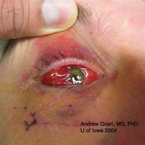
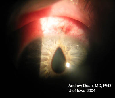
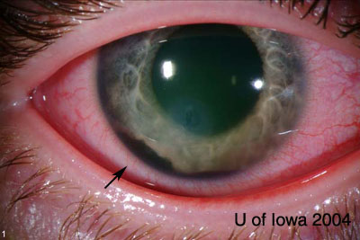
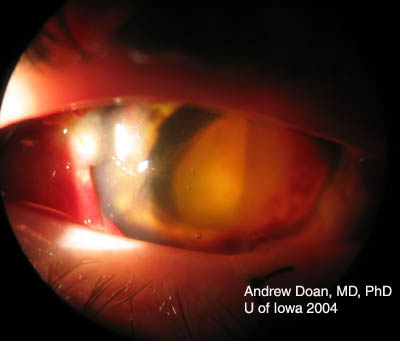
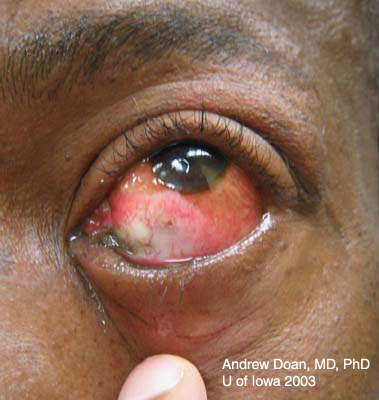
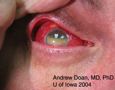
Base injuries are more worrisome as they can penetrate deeper into ocular tissue. Grading of corneal burns is based on extent of limbal ischemia, which indicates loss of corneal epithelial stem cells, and degree of corneal haze obscurring iris details.

Hughes Classification of Ocular Burns

Grade 1 (Very good prognosis)

  • No corneal opacity nor limbal ischemia.

Grade 2 (Good prognosis)

  • Corneal haze but iris details are clear.
  • Less than 1/3 cornea limbus ischemia.

Grade 3 (Guarded prognosis)

  • Sufficient corneal haze to obscure iris details.
  • 1/3 to 1/2 of cornea limbus ischemia.

Grade 4 (Poor prognosis)

  • Opaque cornea without view of iris or pupil.
  • More than 1/2 of cornea limbus ischemia.
Alkali Burn
Alkali Burn
The patient had an ocular burn due to contact with concentrated anhydrous ammonia in an industrial accident. Note the necrotic tissue, small area of inferior limbal ischemia, and epithelial defect of the cornea. There was good iris and pupil details on examination. This was a grade 2 ocular burn.
ENDOPHTHALMITIS
ENDOPHTHALMITIS
A patient with endophthalmitis presenting with decreased vision, pain, redness, and layered white blood cells in the anterior chamber (hypopyon).

MANAGEMENT OF CHEMICAL BURNS

Grading helps to determine the aggressiveness and course of treatment. Treatment modalities include some or all of the following depending on the severity of the burn:

  • Debride necrotic tissue
  • Bandage contact lens
  • Quinolone: 1 drop 4-6x/day (prevents infection)
  • Prednisolone phosphate: 1 drop every 1-2 hr while awake to reduce inflammation
  • Vitamin C: 1-2 gm orally daily to reduce corneal thinning/ulceration
  • 10% sodium citrate: 1 drop every 2 hr while awake (chelates calcium and impairs polymorphonuclear leukocyte (PMN) chemotaxis)
  • Scopolamine 0.25%: 1 drop three times daily (reduces pain/scarring with anterior chamber inflammation)
  • 10% Mucomyst® (n-acetyl-cysteine): 1 drop 6x/day (mucolytic agent and collagenase inhibitor)
  • Doxycycline 100 mg orally twice daily (collagenase inhibitor)
  • Glaucoma drops/oral acetazolamide (Diamox®) if IOP elevated
  • Significant injury may require admission

ENDOPHTHALMITIS

The Endophthalmitis Vitrectomy Study (EVS) [2] applies only to post-cataract endophthalmitis. The study states that patients did better with a vitrectomy and injection of intravitreal antibiotics when the vision is light perception or worse. If vision is hand motions or better, then patients with a TAP (vitreous biopsy and culture) and intravitreal injection of antibiotics did as well as patients who had a vitrectomy and injection of intravitreal antibiotics.

If referral doctor suspects endophthalmitis, ask about penicillin allergy before patient is enroute.

  • Order intravitreal antibiotics immediately.
  • Call the senior resident so they can help you set up the minor room for a tap and inject. This can be difficult if the patient does not have a hospital number. In the past, I have given the patient's name and birthdate to the pharmacist, and they can look up the patient after they check in. The intravitreal injections only have a shelf life of about 2 hours, so do not order them too early.
  • Obtain gram stain and culture media (plates & liquid culture media) for aqueous and vitreous (1 set for each).

INTRAVITREAL ANTIMICROBIAL CONCENTRATIONS

  • Intravitreal vancomycin: 1 mg/0.1 ml in 1 cc syringe.
  • Intravitreal ceftazidime: 2.25 mg/0.1 ml in 1 cc syringe.
  • Penicillin Allergy: Intravitreal gentamycin: 100 µg/0.1 ml in 1 cc syringe or amikacin 200-400 µg/0.1 ml in 1 cc syringe. Some attending physicians prefer amikacin, due to its somewhat better toxicity profile.
  • Be sure there is at least 0.5 ml of antibiotic in syringe so you can transfer to a sterile syringe and still have enough left for your injection.
  • Suspect Bacillus cereus: Intravitreal clindamycin: 0.5 mg/0.1 ml (optional). To date, B. cereus is sensitive to Vancomycin.
  • Suspect fungus: Intravitreal amphotericin B: 5-10 µg/0.1 ml.

References

  1. Nerad, JA. Oculoplastic Surgery. The Requisites in Ophthalmology. Mosby 2001. 348--386.
  2. Results of the Endophthalmitis Vitrectomy Study. A randomized trial of immediate vitrectomy and of intravenous antibiotics for the treatment of postoperative bacterial endophthalmitis. Endophthalmitis Vitrectomy Study Group. Arch Ophthalmol. 1995. Dec; 113 (12): 1479-96.
  3. Thach, Allen B. Ophthalmic Care of the Combat Casualty. Textbook of the Military Medicine Series. Office of The Surgeon General, United States Army. June 1, 2003.
  4. Virtual Naval Hospital. Ocular Trauma Manual. http://www.vnh.org/Providers.html (noted on review, Jan 2011, this resource has been discontinued by the US Government, an archived copy may be found at http://web.archive.org)
  5. Wagoner, MD. Chemical injuries of the eye: current concepts in pathophysiology and therapy. Surv Ophthalmol. 1997 Jan-Feb; 41 (4): 275-313.