73-year-old male, with no prior past ocular history, presented at the referral of an optometrist for the possibility of retinoschisis in both eyes. He was asymptomatic without any flashes, floaters, or peripheral vision loss. He had no family history of any retinal problems.
BCVA: OD 20/20, OS 20/30 with correction
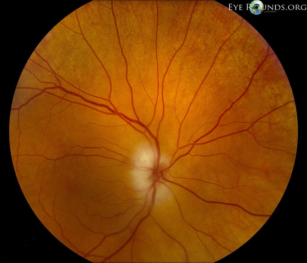
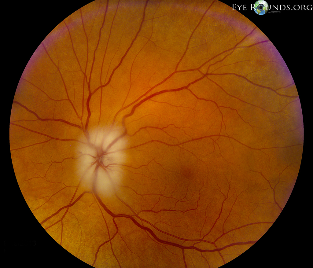
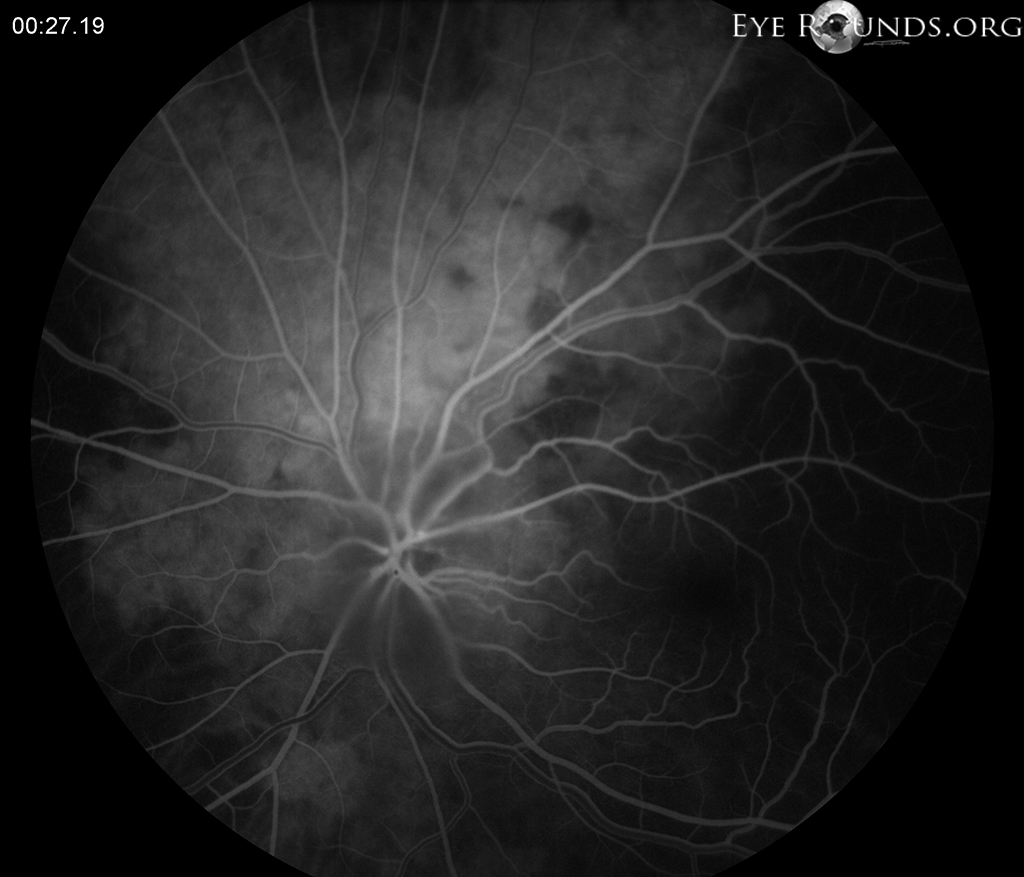
SLE: 3+ NS OU; no PVD OU DFE: normal without any peripheral retinal tears, holes, or detachments; inferotemporally there was a dome-shaped 'boggy-area' of elevated retina near the retina in both eyes. B-scan OU: mild vitreous opacities, dome-shaped retinal elevation inferotemporally consistent with retinoschisis.
Ophthalmic Atlas Images by EyeRounds.org, The University of Iowa are licensed under a Creative Commons Attribution-NonCommercial-NoDerivs 3.0 Unported License.


Address
University of IowaLegal
Related Links