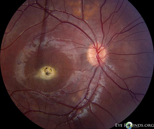
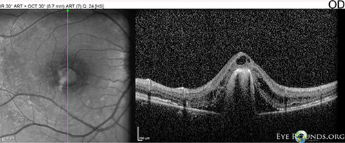
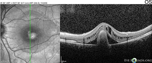
These are photographs from a 6-year-old boy with no known past medical history who presented for decreased visual acuity in both eyes (OU) that was first noticed when he failed his preschool vision screening at age four. His visual acuity with correction was 20/200+2 OD and 20/80+1 OS, without pinhole improvement. Confrontational visual fields, intraocular pressure, extraocular movements, and slit lamp exam were all unremarkable. On dilated fundus exam,there was a large (slightly less than 1 disc diameter) white elevated fibrotic scar in the fovea with overlying pigmentation and surrounding edema OU. The near periphery demonstrated the extrafoveal yellow-colored vitelliform lesions that are characteristic of autosomal recessive Best disease. These likely represent lipofuscin accumulation. Optical coherence tomography showed a large fibrovascular pigment epithelial detachment in each fovea (due to chronic inactive choroidal neovascular membranes OU), associated with parafoveal subretinal fluid, and overlying cystic retinal edema OU. There was loss of the inner segment/outer segment junction in this same area centrally OU. Autosomal recessive Best disease is usually caused by two "mild" mutations in the BEST1 gene.

Ophthalmic Atlas Images by EyeRounds.org, The University of Iowa are licensed under a Creative Commons Attribution-NonCommercial-NoDerivs 3.0 Unported License.


Address
University of IowaLegal
Related Links