This site uses tracking information. Visit our privacy policy. Click to agree to this policy and not see this again.

Ophthalmology and Visual Sciences

Corneal Hydrops

Corneal Hydrops

Category(ies): Cornea, External Disease
Contributor: Jesse Vislisel, MD
Photographer: Jody Troyer

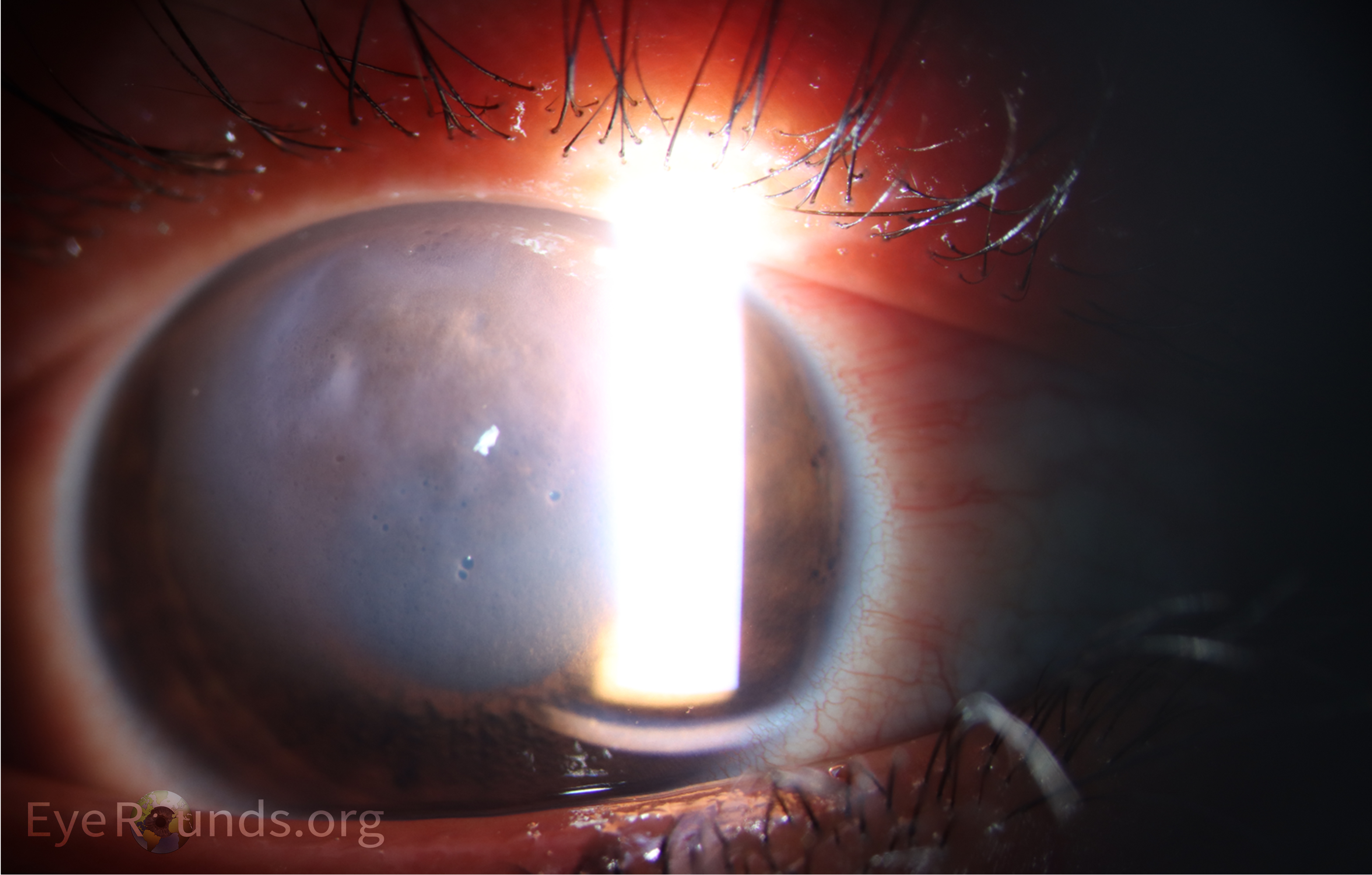
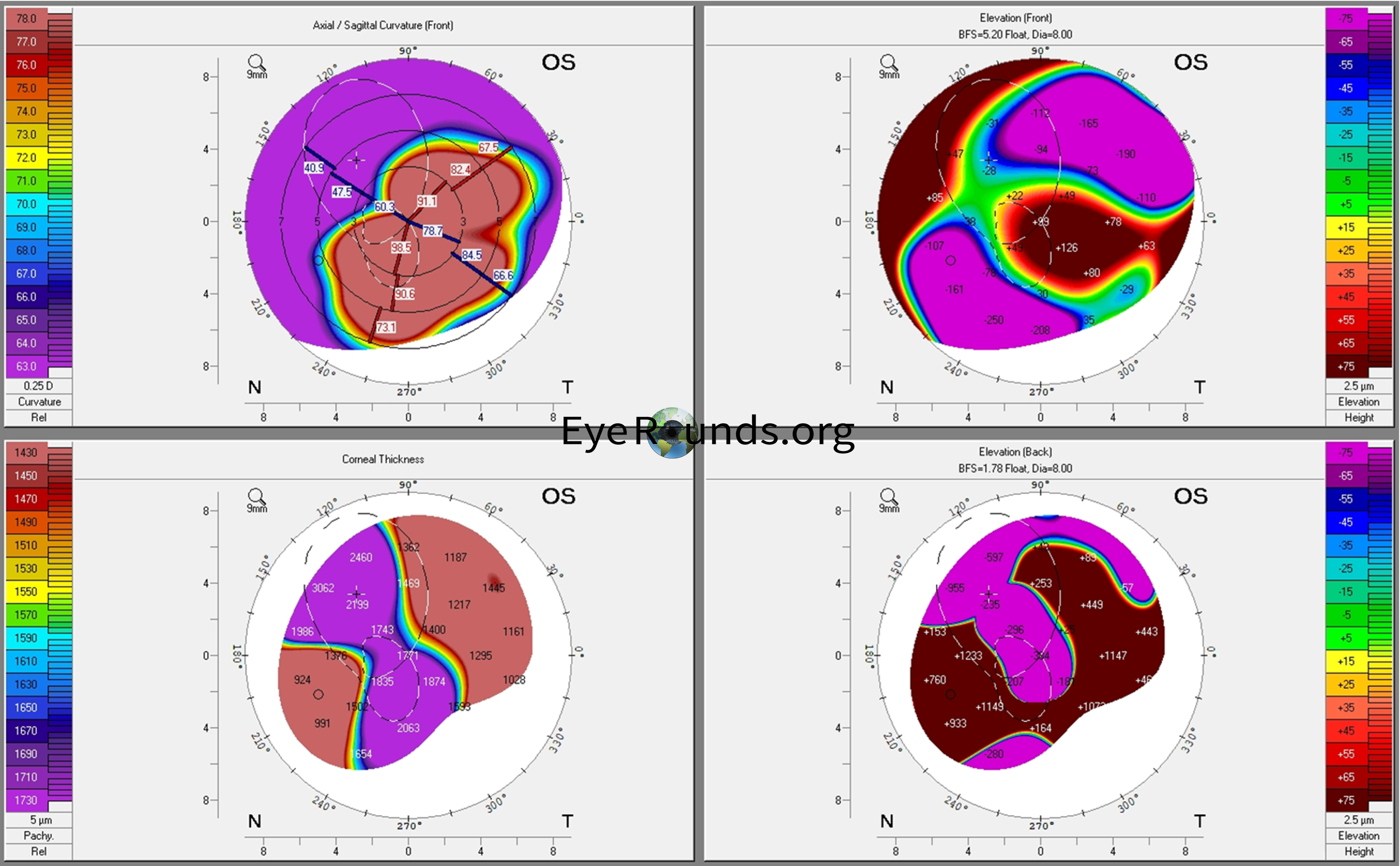
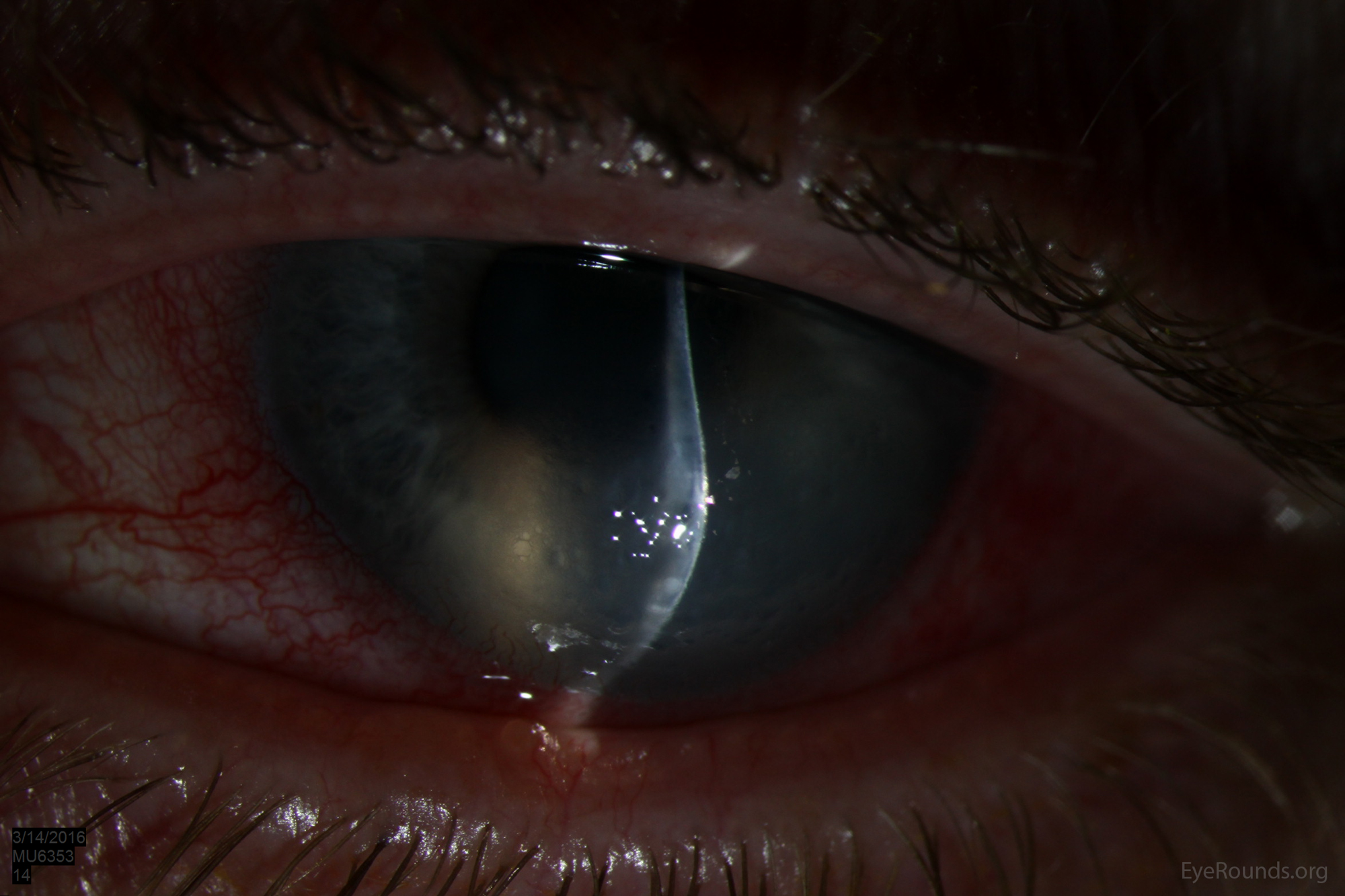
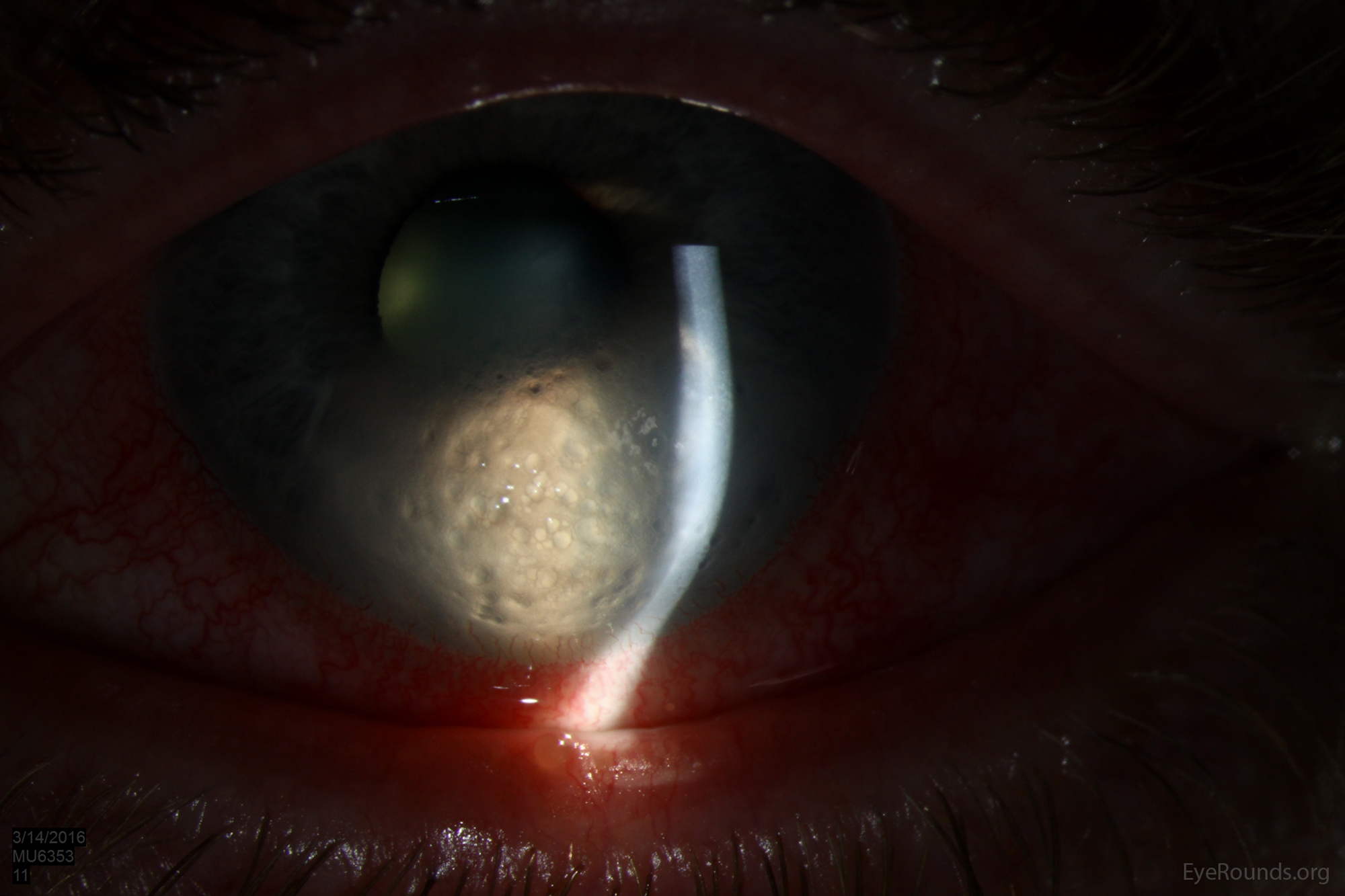
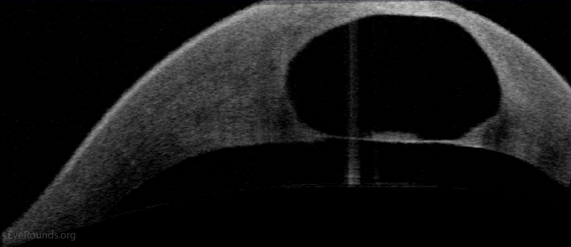
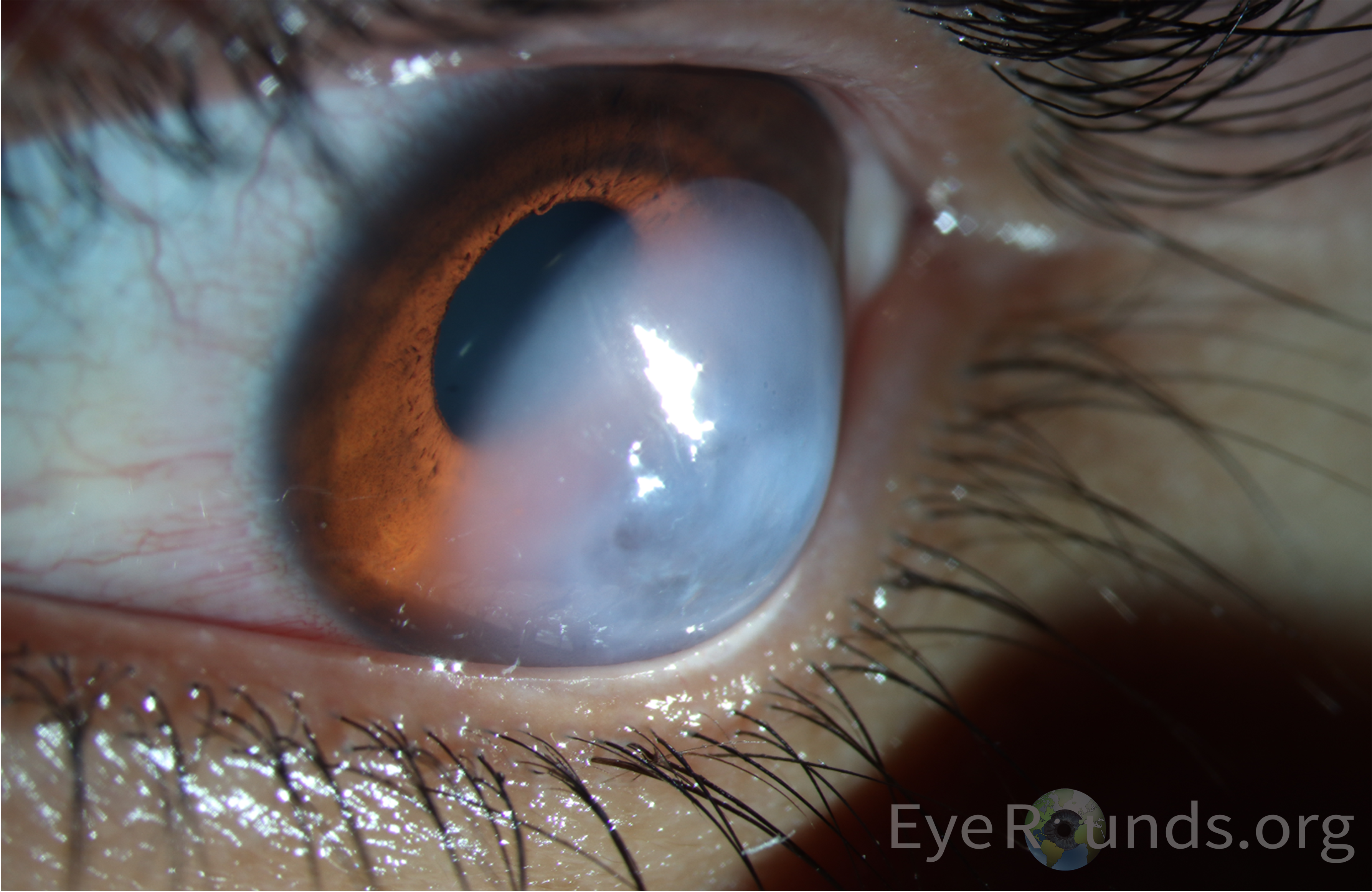
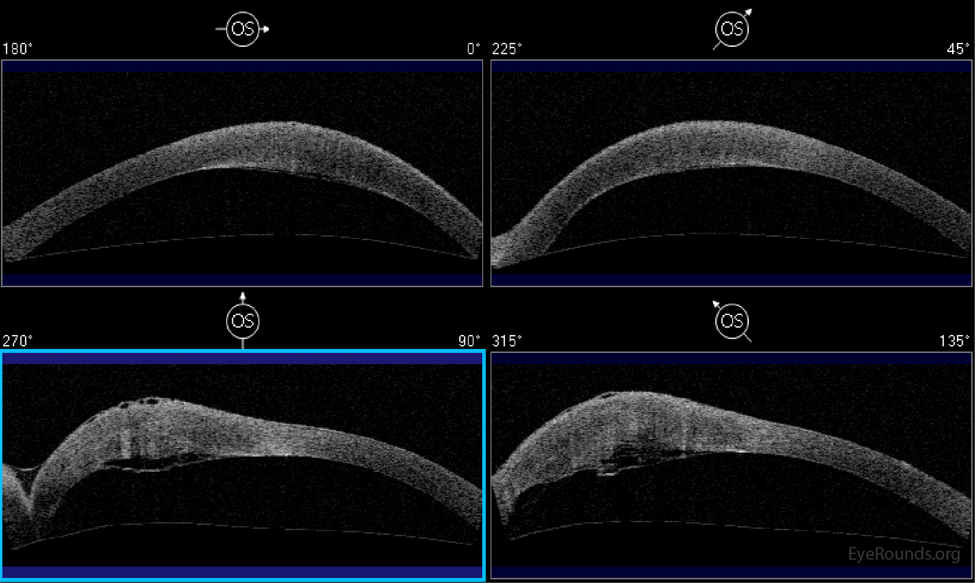
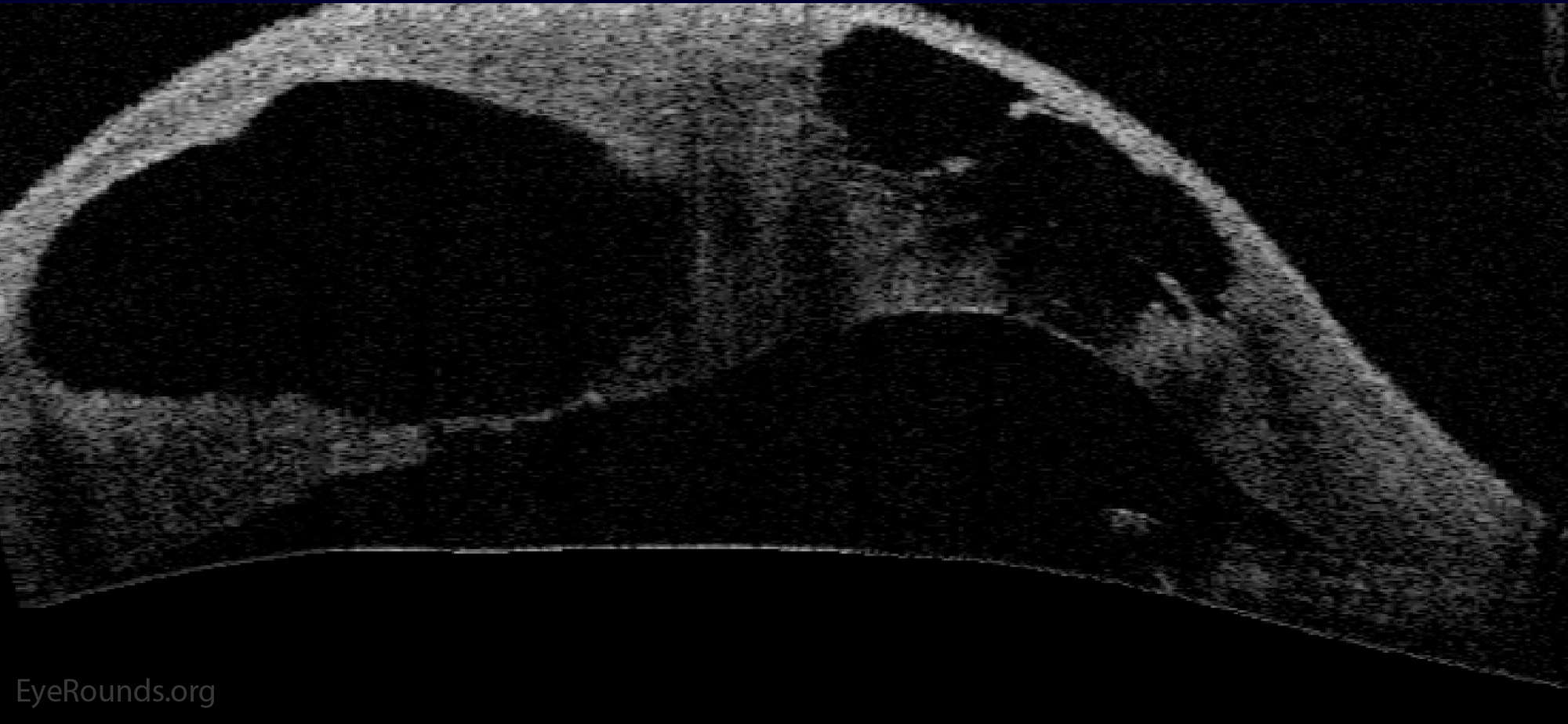
Corneal hydrops is the acute onset of corneal edema due to a break in Descemet membrane. This condition may be seen in individuals with advanced keratoconus or other forms of corneal ectasia. Recovery may take weeks to months with medical therapy, but may be accelerated by the placement of air or gas into the anterior chamber to slow the influx of aqueous into the cornea. Scarring, and sometimes corneal flattening, will occur after resolution of the episode.


Entry #1

Entry #1

Contributor: Matthew Meyer, MD
Photographer: Sarah Skiles
February 12, 2024

Oblique slit-lamp view of acute corneal hydrops highlighting significant apical corneal edema with corresponding anterior slit-lamp view and corneal tomography.

Oblique slit-lamp view of acute corneal hydrops highlighting significant apical corneal edema
Fig 1. Oblique slit-lamp view of acute corneal hydrops highlighting significant apical corneal edema
Anterior slit-lamp view of acute corneal hydrops highlighting significant apical corneal edema
Fig 2. Anterior slit-lamp view
Corneal tomography of acute corneal hydrops highlighting significant apical corneal edema
Fig 3. Corneal tomography

Entry #2

Entry #2

Contributor: Jesse Vislisel, MD
Photographer: Jody Troyer
acute hydrops, sever inferior corneal stromal edema
Fig 1a. Acute corneal hydrops with severe inferior corneal stromal edema and overlying microcystic edema and bullae in the setting of advanced keratoconus. These photographs were taken 1 week after onset of the episode.
acute hydrops, sever inferior corneal stromal edema slit lamp
Fig 1b. Slit Lamp
acute hydrops, sever inferior corneal stromal edema wide slit lamp
Fig 1c. Wide Slit Lamp
OCT showing stromal edema
Fig 2. Anterior segment OCT of the patient in Figure 1 demonstrating the severe stromal edema in the area of involvement with underlying detachment of Descemet membrane in the area of rupture. Epithelial bullae can be seen overlying the area of stromal edema.
OCT showing stromal edema
Fig 3a. Anterior segment OCT of a patient with keratoconus and severe corneal hydrops. These images demonstrate the large, cystic accumulations of intrastromal fluid that were present. Subsequent penetrating keratoplasty was required.
OCT showing stromal edema zoomed
Fig 3b. Anterior segment OCT of a patient with keratoconus and severe corneal hydrops. These images demonstrate the large, cystic accumulations of intrastromal fluid that were present. Subsequent penetrating keratoplasty was required.