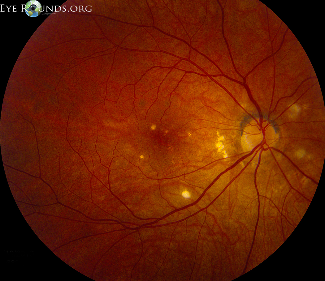
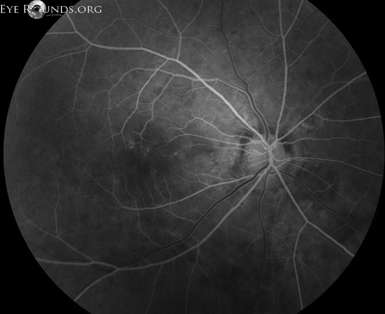
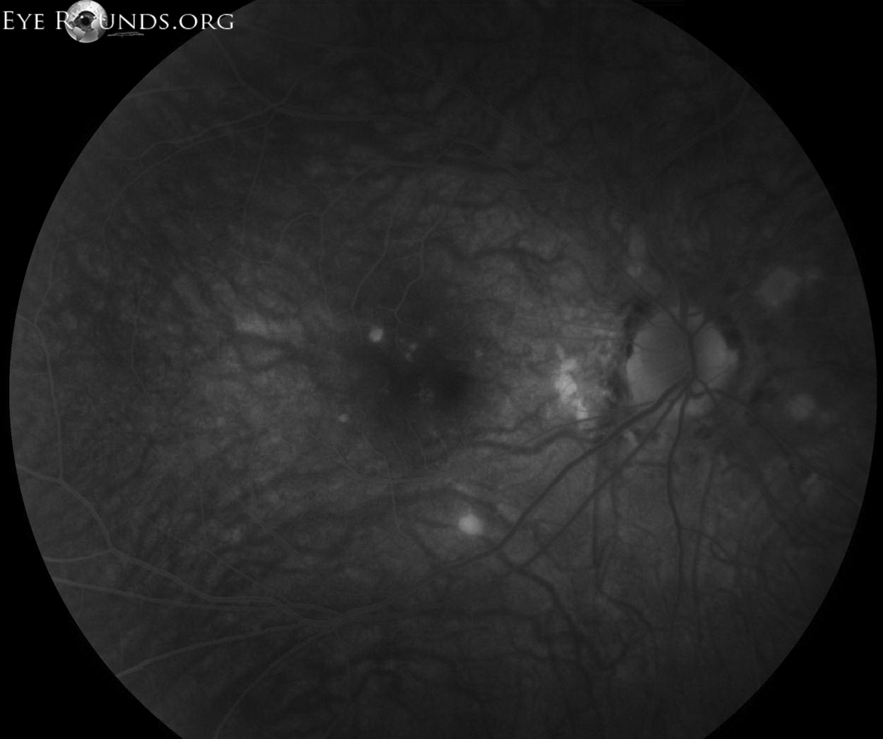
Punctate inner choroiditis/choroidopathy (PIC) is an inflammatory disorder most common in otherwise healthy, young, white, myopic women. The condition results in small (100-200 µm) yellow-white chorioretinal lesions in the posterior pole, rarely extending to the midperiphery. The lesions progress to deep, punched-out chorioretinal scars, often with surrounding hyperpigmentation. In contrast to some similar conditions, there is no associated vitritis. Fluorescein angiography shows early hyperfluorescence and late staining of the lesions, as seen in these photographs. The disease course is self-limited and there is generally a good visual prognosis, though vision can be limited in the presence of choroidal neovascularization.

Ophthalmic Atlas Images by EyeRounds.org, The University of Iowa are licensed under a Creative Commons Attribution-NonCommercial-NoDerivs 3.0 Unported License.

Address
University of IowaLegal
Related Links