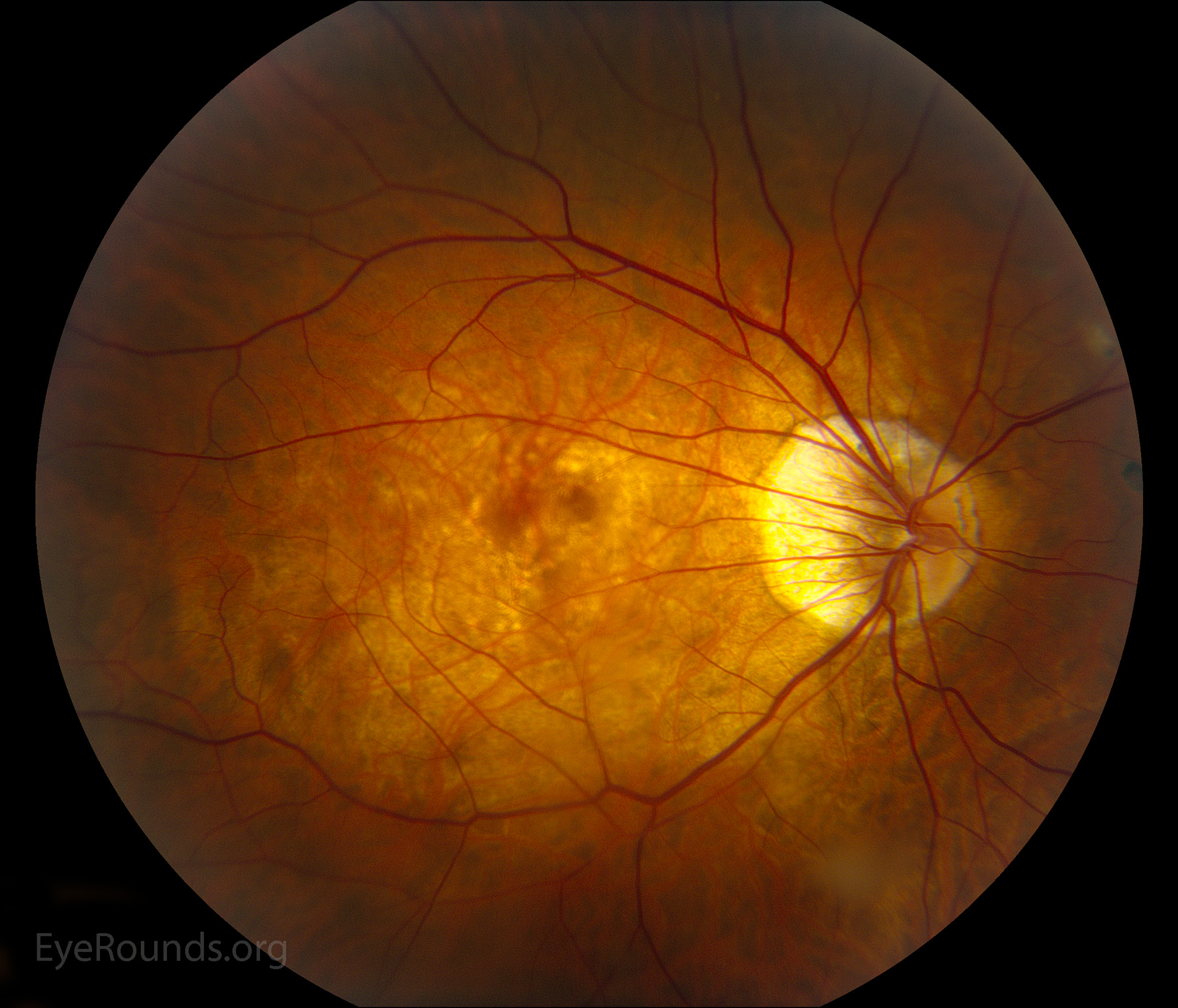
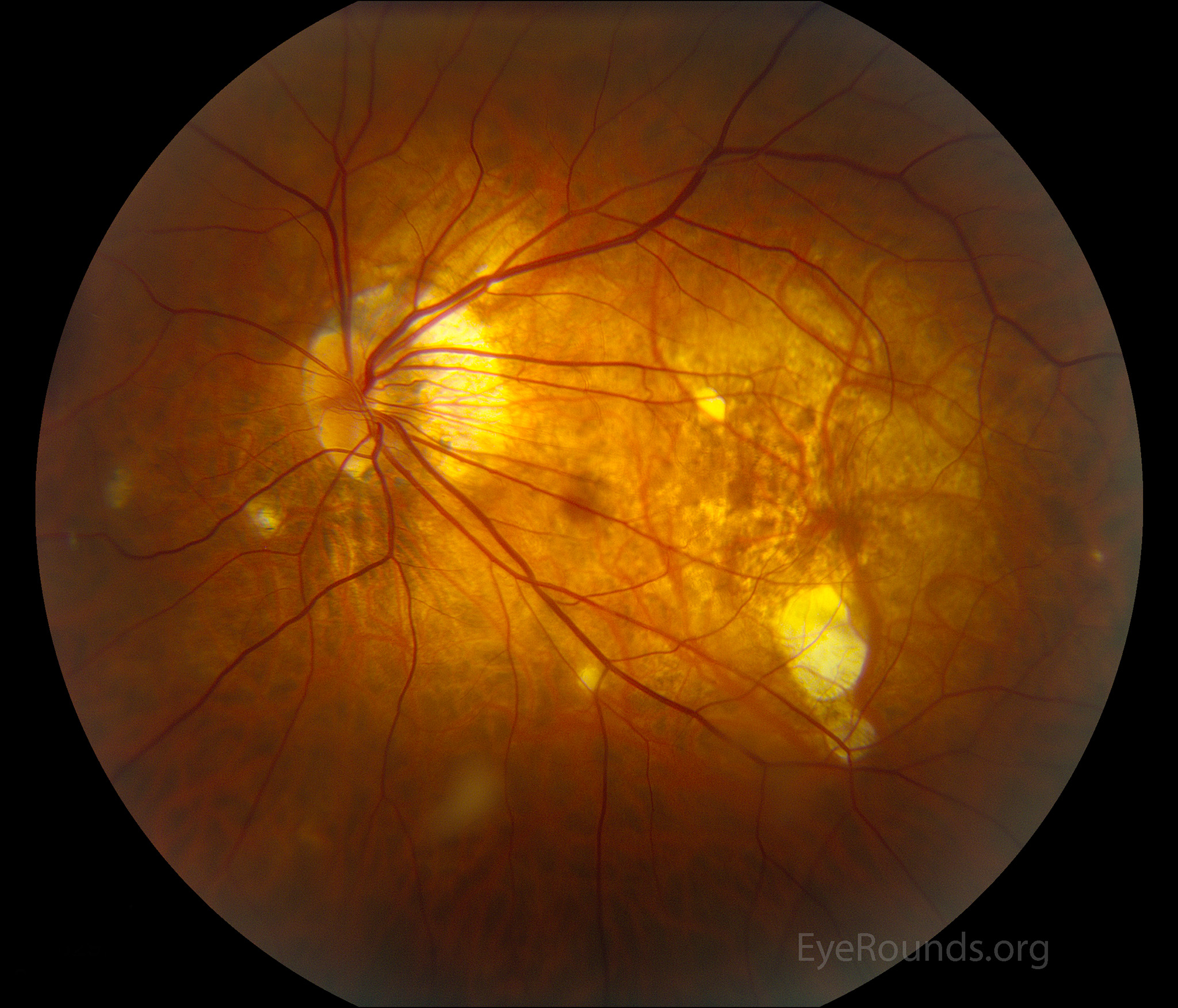
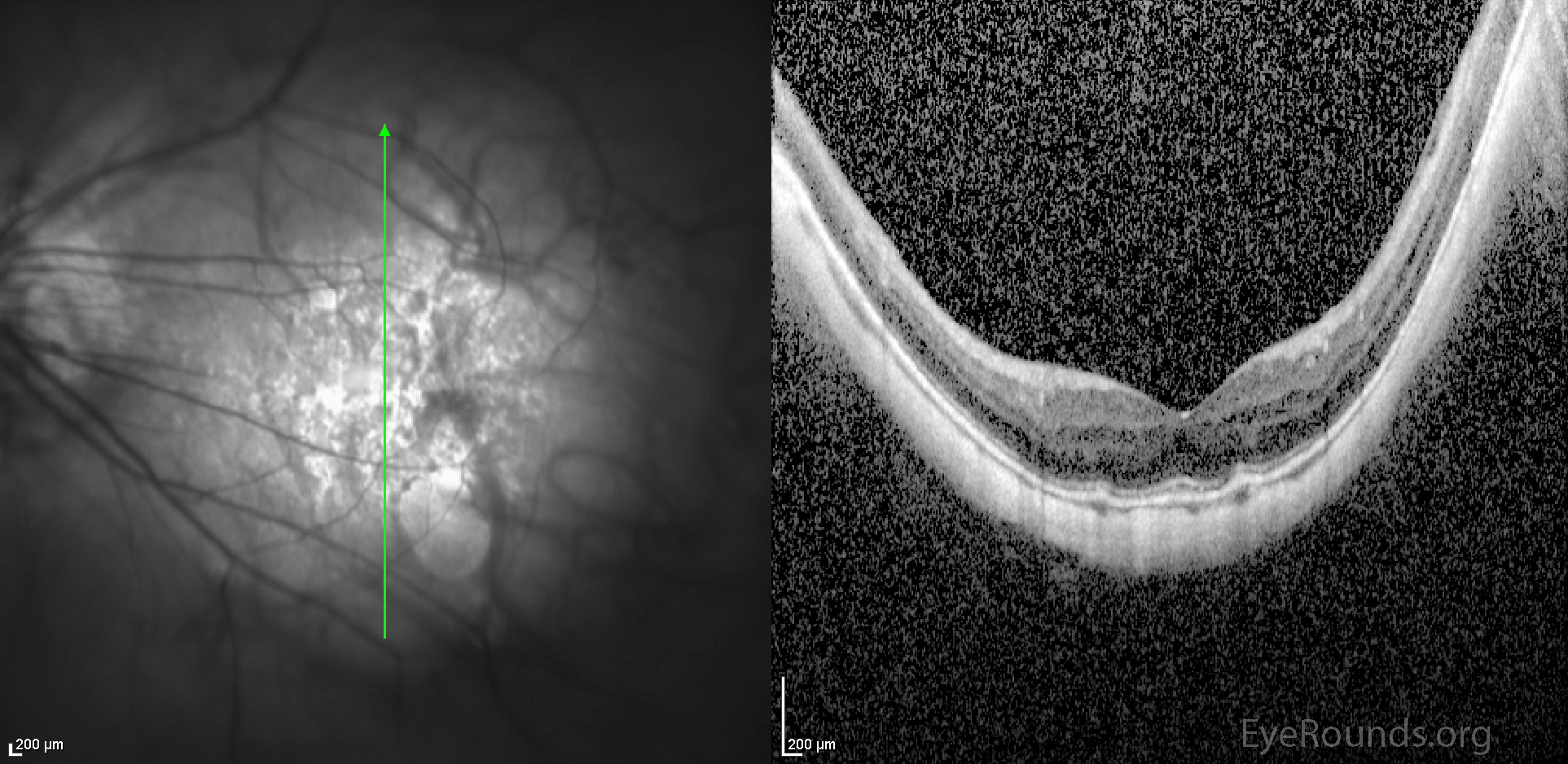
Pathologic myopia is defined as myopia over -8.00 D or an axial eye length >32.5 mm. These eyes may undergo progressive elongation with development of degenerative changes of the retina, retinal pigmented epithelium (RPE), and choroid. These photographs show the fundus findings in two eyes with a spherical prescription of -17.00 D. Note the tilted optic discs, peripapillary atrophy, central macular RPE thinning with several discrete regions of RPE atrophy in the left eye, and bilateral posterior staphylomas (the central maculae appear blurred as they are out of the plane of focus). The OCT images show the staphylomas more clearly as a bowing of the posterior structures of the eye.

Ophthalmic Atlas Images by EyeRounds.org, The University of Iowa are licensed under a Creative Commons Attribution-NonCommercial-NoDerivs 3.0 Unported License.

Address
University of IowaLegal
Related Links