This site uses tracking information. Visit our privacy policy. Click to agree to this policy and not see this again.

Ophthalmology and Visual Sciences

Pigmented Conjunctival Nevus

Pigmented Conjunctival Nevus

Category(ies): Cornea, External Disease
Contributor: Dr Rajan Paul


Conjunctival nevus occurring in childhood

Conjunctival nevus occurring in childhood.

Category(ies): Cornea / External Eye Disease
Contributor: Ella J. Gehrke, MD; Kanwal Singh Matharu, MD; Pavlina Kemp, MD
Photographer: Michael T. Edrington, CRA
Posted: 03/03/2025

A 
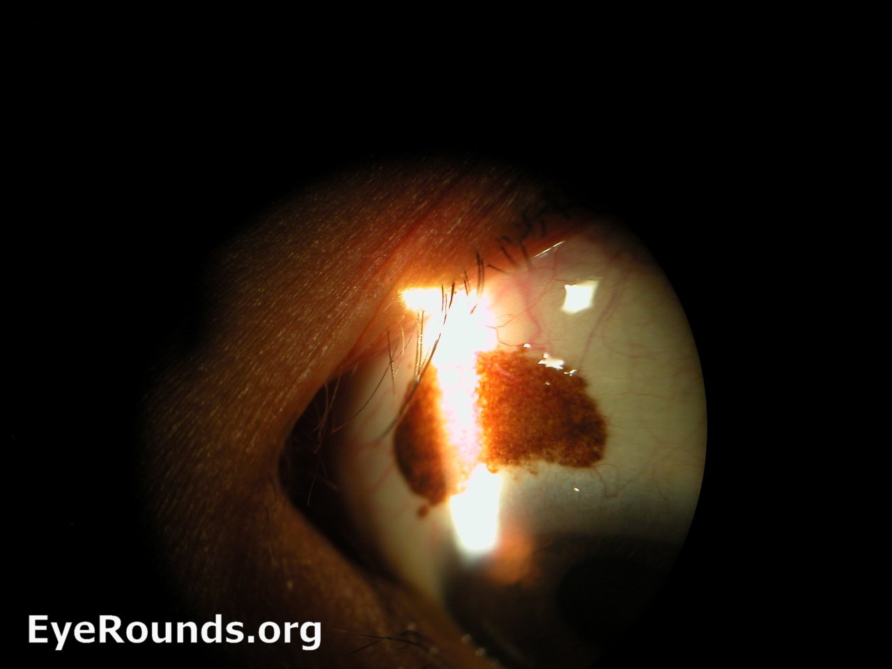
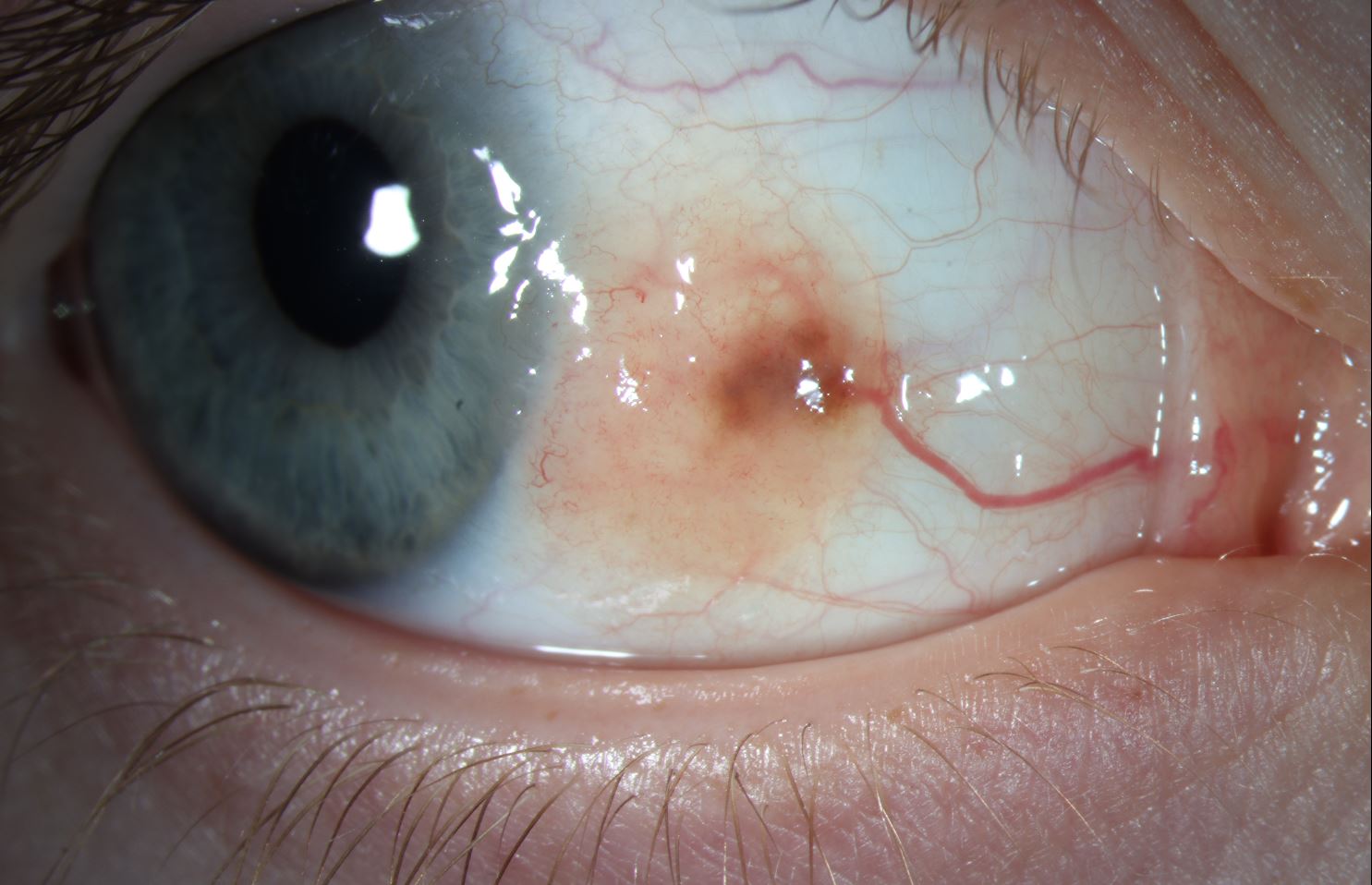
10-year-old female presented with a conjunctival lesion at the nasal limbus of her right eye. The lesion measured approximately 1 cm × 6.5 mm, with notable elevation and a melanotic core. A prominent feeder vessel was also observed. The lesion had been slowly enlarging over six years. Her visual acuity was 20/20 OU, with no reported diplopia, and intraocular pressures were within normal limits. Surgical excision was performed, and histopathology confirmed a juvenile compound nevus.

Elevated, irregularly shaped, salmon-colored conjunctival lesion measuring 1 cm × 6.5 mm, with a faint melanotic core and a large feeder vessel, located at the nasal limbus of the right eye.

Image Permissions:
Creative Commons

Ophthalmic Atlas Images by EyeRounds.org, The University of Iowa are licensed under a Creative Commons Attribution-NonCommercial-NoDerivs 3.0 Unported License.