This site uses tracking information. Visit our privacy policy. Click to agree to this policy and not see this again.

Ophthalmology and Visual Sciences

Retinal detachment with proliferative vitreoretinopathy

Retinal detachment with proliferative vitreoretinopathy

Category(ies): Retina, Vitreous
Contributor: Jesse Vislisel, MD
Photographer: Brice Critser, CRA

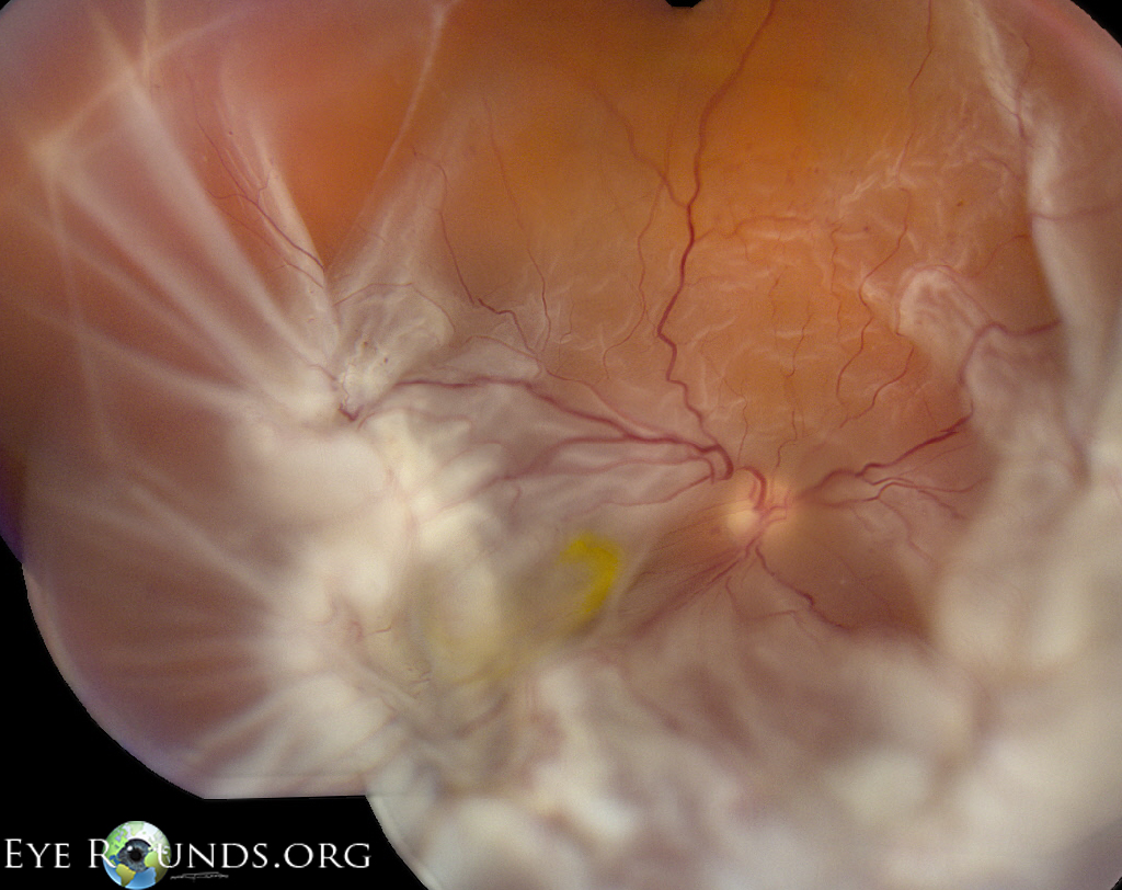
Proliferative vitreoretinopathy (PVR) is a condition characterized by tractional retinal membranes formed by glial, retinal pigmented epithelial, and other cells. It is the most common cause for failure of retinal detachment repair. These contractile membranes result in generalized retinal shrinkage, fixed retinal folds, and characteristic "star folds," which are most evident from 4:00 to 11:00 in this photograph of a retinal detachment involving the macula.


Retinal detachment with proliferative vitreoretinopathy

Retinal detachment with proliferative vitreoretinopathy

Category(ies): Retina, Vitreous
Contributor: Alina Husain, MD; Justin Grassmeyer, MD, PhD; Elaine M. Binkley, MD; Ian Han, MD
Photographer: Alexandria Steggall

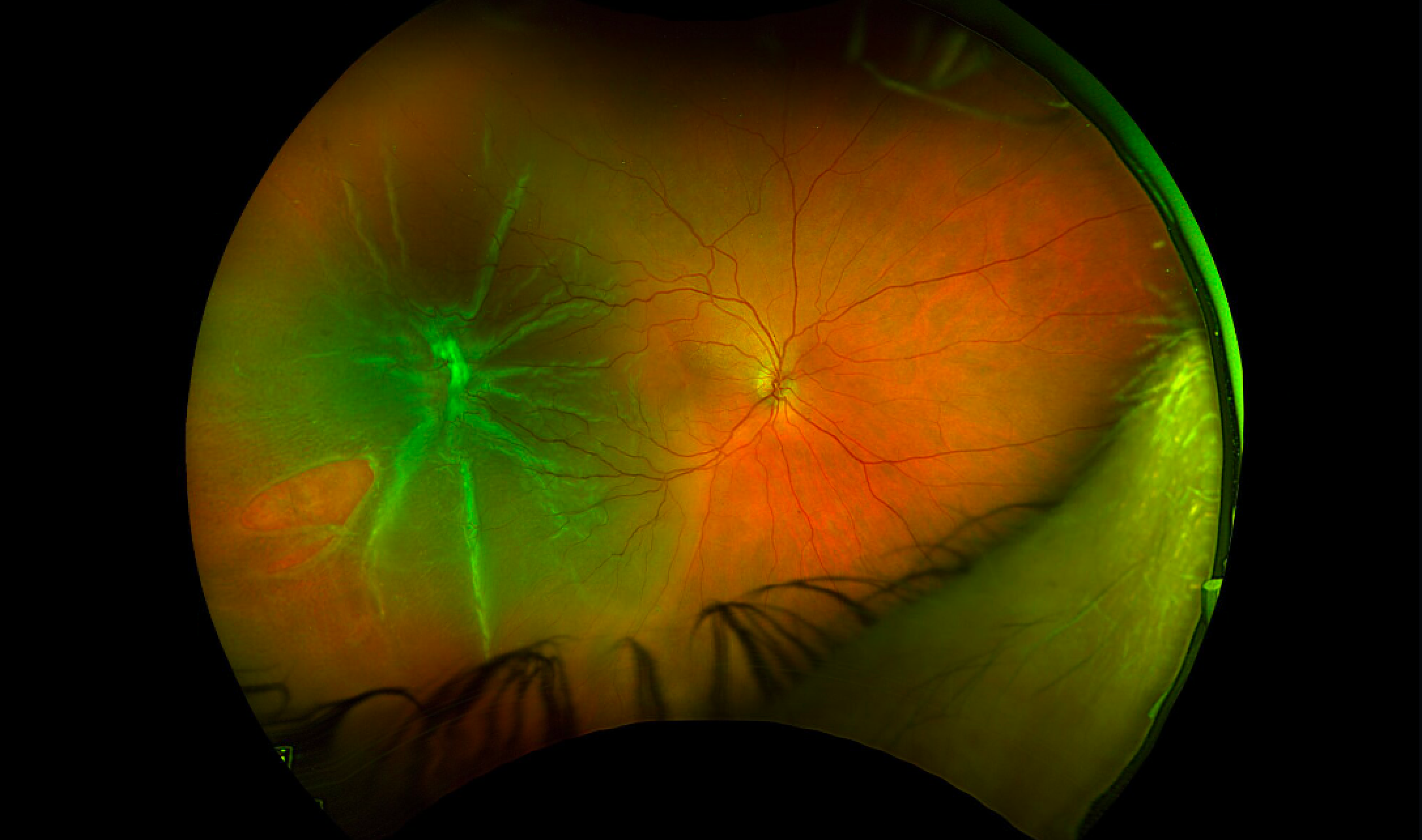
A 66-year-old female was found to have an acute-on-chronic macula-involving rhegmatogenous retinal detachment with Grade C proliferative vitreoretinopathy. The patient underwent retinal detachment repair utilizing 25-gauge vitrectomy with scleral buckle, endolaser, and 14% C3F8 tamponade. The preretinal membranes were peeled during surgery, resulting in relaxation of traction and retinal re-attachment.

Wide-field pseudocolor fundus photograph of the right eye shows a macula-involving rhegmatogenous retinal detachment associated with a prominent star fold temporally and a retinal tear in the inferotemporal periphery
Figure 1. Wide-field pseudocolor fundus photograph of the right eye shows a macula-involving rhegmatogenous retinal detachment associated with a prominent star fold temporally and a retinal tear in the inferotemporal periphery

Retinal detachment with proliferative vitreoretinopathy

Retinal detachment with proliferative vitreoretinopathy

Category(ies): Retina, Vitreous
Contributor: Alina Husain, MD; Justin Grassmeyer, MD, PhD; Elaine M. Binkley MD

This patient is a 68-year-old male who presented with a macula-involving rhegmatogenous retinal detachment of the left eye due to rapid and aggressive development of proliferative vitreoretinopathy. Within the preceding month, the patient had undergone laser demarcation of a superior retinal tear, pars plana vitrectomy for vitreous hemorrhage, and laser demarcation of a localized inferior retinal detachment. He subsequently underwent retinal detachment repair utilizing 25-gauge pars plana vitrectomy, scleral buckle, inferior retinectomy, and silicone oil tamponade.
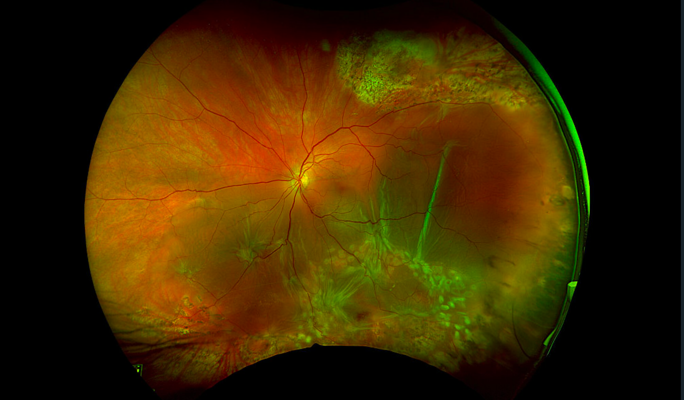

 Wide-field pseudocolor fundus photograph, left eye. There is a macula-involving retinal detachment due to inferior proliferative vitreoretinopathy with multiple star folds. There are laser scars from barricade of a prior retinal tear (superiorly) and detachment (inferiorly).
Figure 1. Wide-field pseudocolor fundus photograph, left eye. There is a macula-involving retinal detachment due to inferior proliferative vitreoretinopathy with multiple star folds. There are laser scars from barricade of a prior retinal tear (superiorly) and detachment (inferiorly).