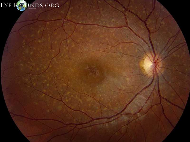
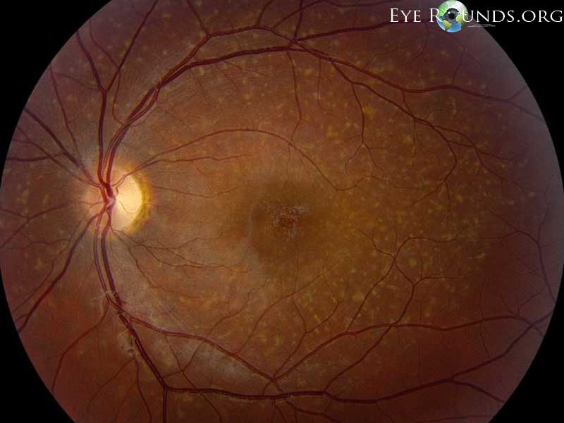
20-year-old male with molecularly confirmed Stargardt disease (ABCA4 mutation). Note the diffuse yellow pisciform flecks within the arcades and central macular mottling in both eyes.
OMIM #248200, #600110, #603786
Ed Stone, MD, PhD teaches the following:
A photoreceptor cell-specific ATP-binding transporter gene (ABCR) is mutated in recessive Stargardt macular dystrophy.
Most common mutation is Gly1961Glu
Clinical features:
If patients are 20/40, then they'll be 20/200 in 5 years on average.
Patients have exuberant response to incidental ocular trauma- keloid scars in macular. Avoid contact sports.
FFA demonstrates masked choroid.
Heidelberg autofluoresence is present.

Ophthalmic Atlas Images by EyeRounds.org, The University of Iowa are licensed under a Creative Commons Attribution-NonCommercial-NoDerivs 3.0 Unported License.


Address
University of IowaLegal
Related Links