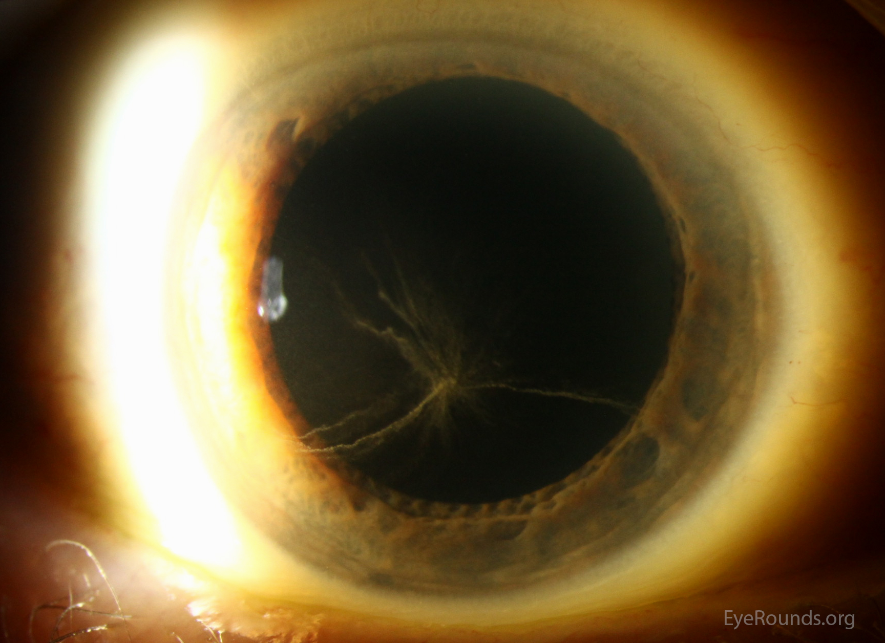
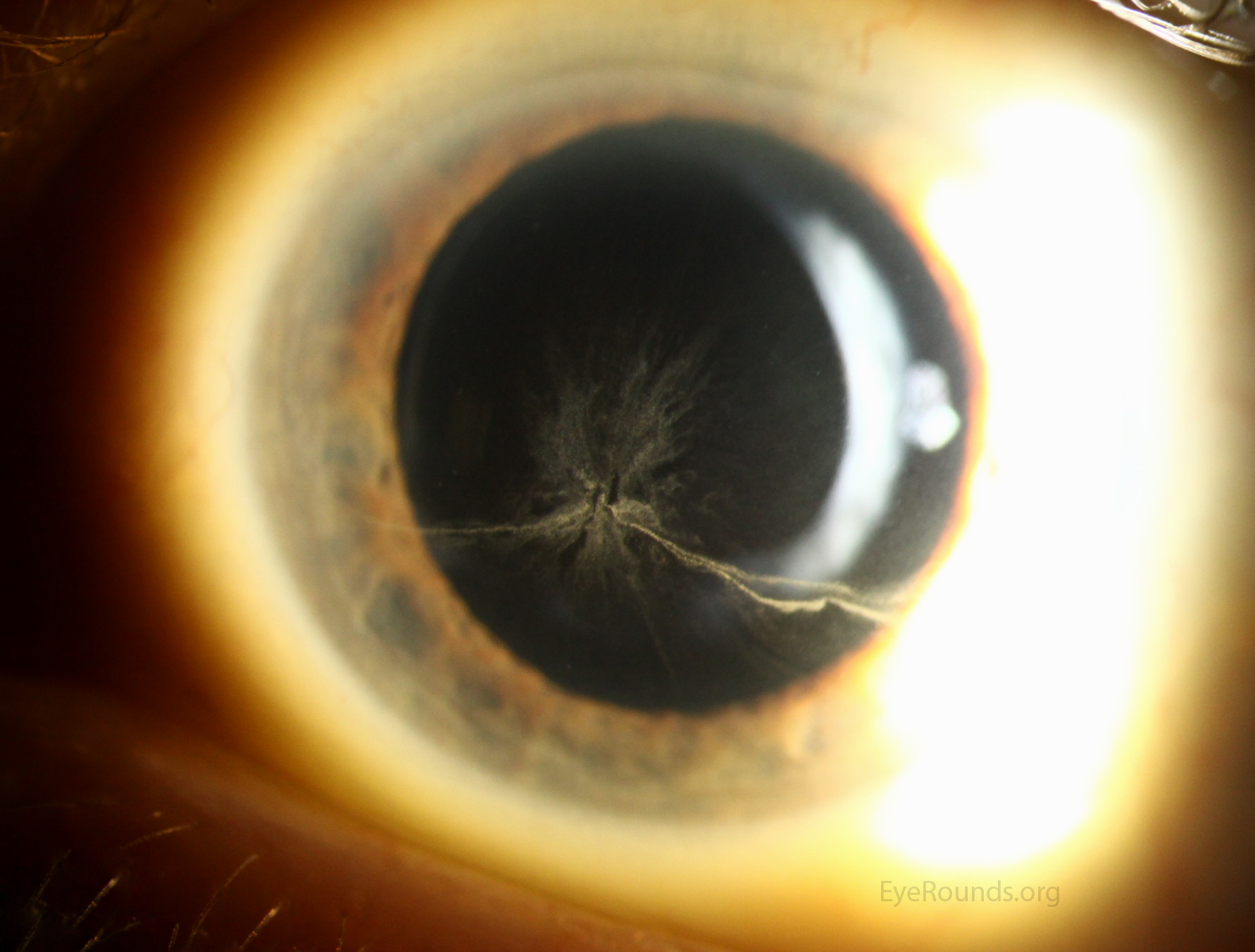
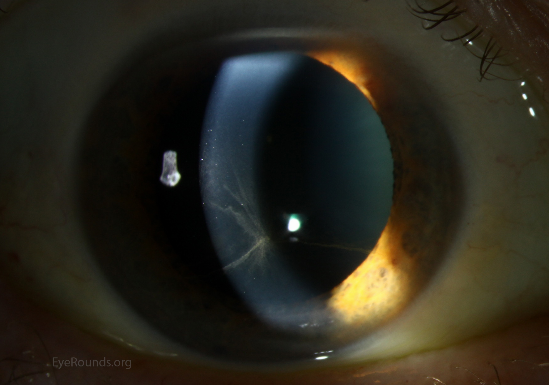
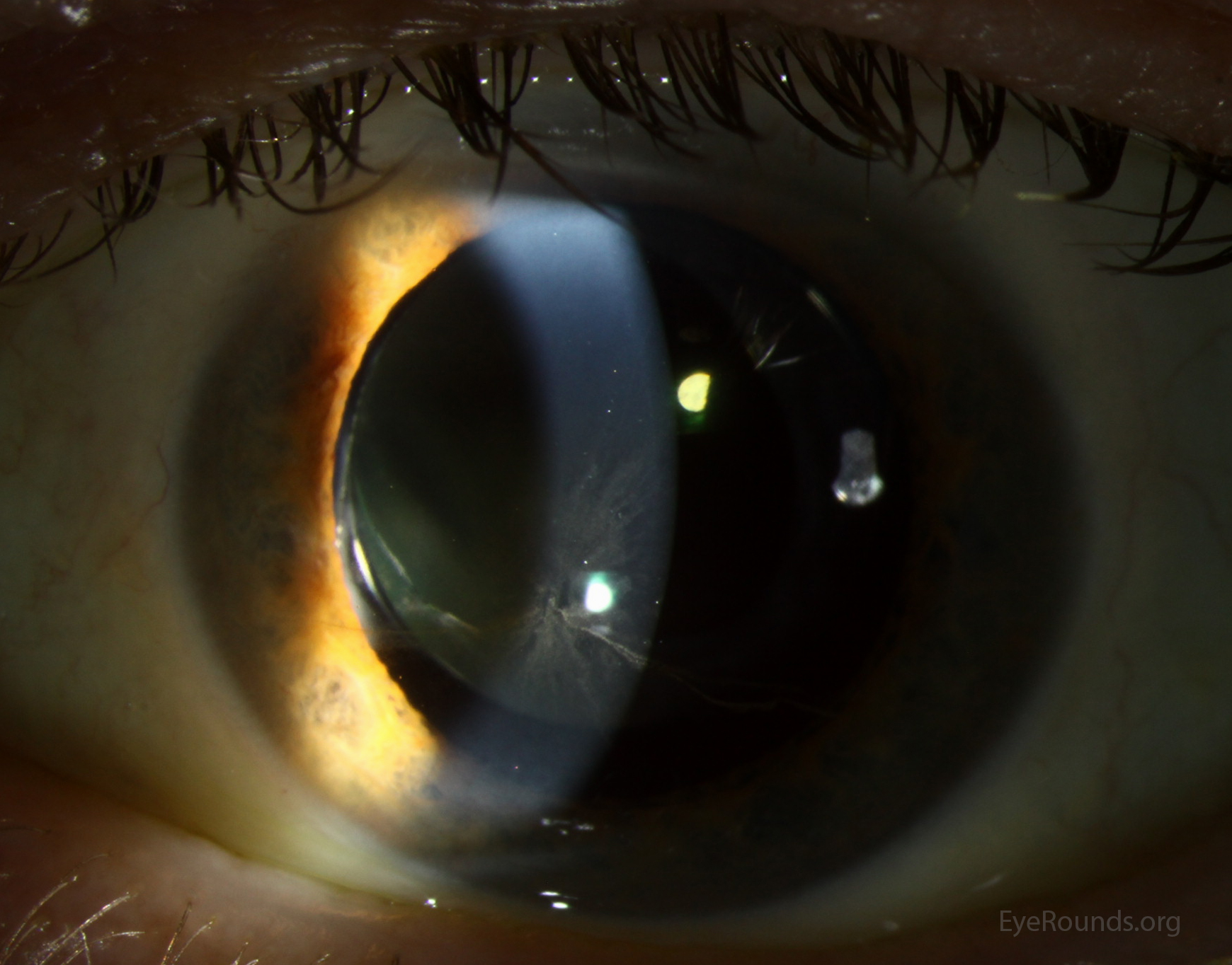
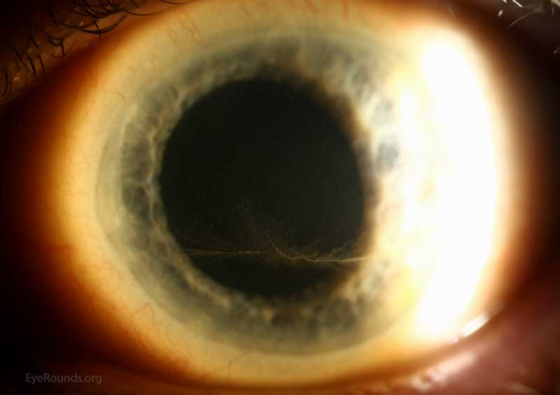
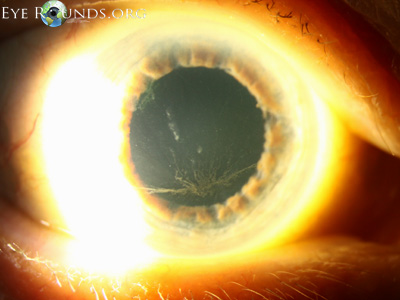
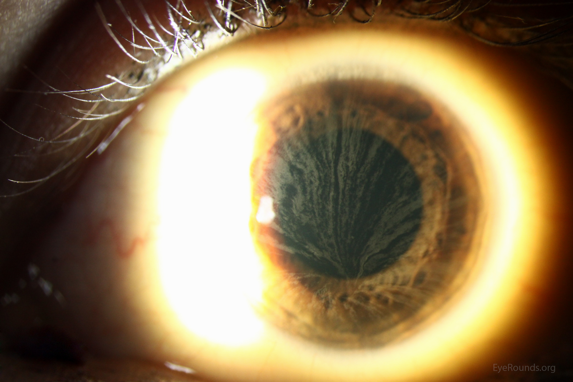
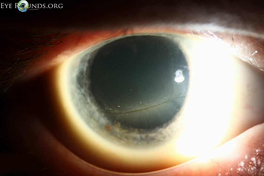
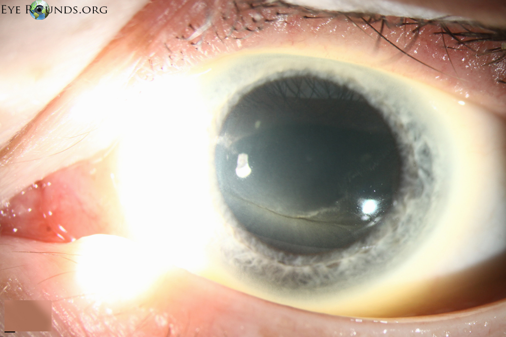
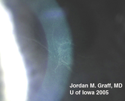
Cornea verticillata, also known as vortex keratopathy, describes a pattern of whorl- shaped opacities within the basal corneal epithelium. They are most commonly located in the inferior paracentral region, are non-elevated, and can range from white to brown in color. These changes are usually not visually significant. Cornea verticillata is often caused by the use of certain systemic medications, the most common of which include amiodarone, chloroquine, hydroxychloroquine, indomethacin, and phenothiazenes. Cornea verticillata can also be seen in the sphingolipidosis, Fabry disease.

Ophthalmic Atlas Images by EyeRounds.org, The University of Iowa are licensed under a Creative Commons Attribution-NonCommercial-NoDerivs 3.0 Unported License.

Address
University of IowaLegal
Related Links