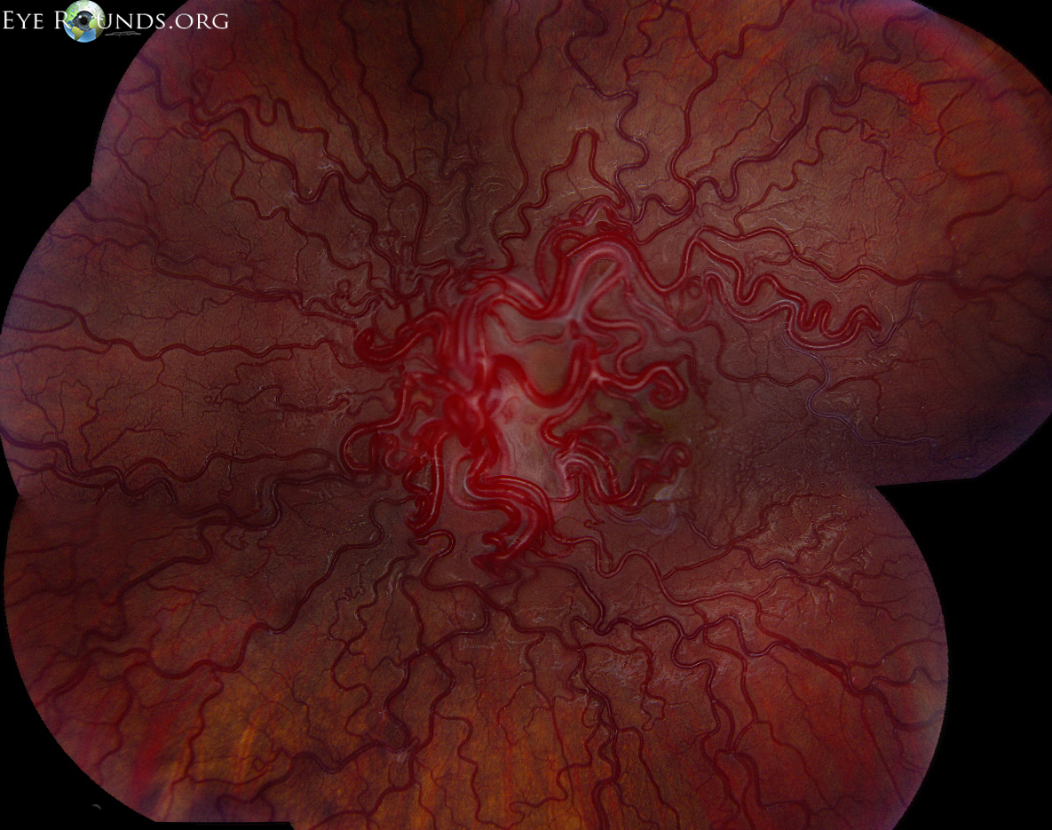
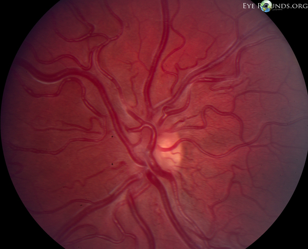
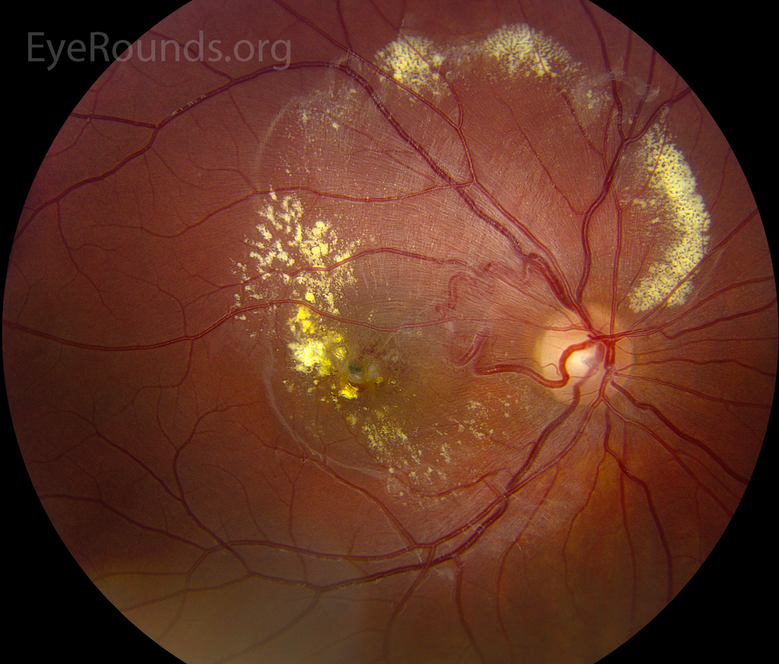
Wyburn-Mason syndrome (racemose hemangiomatosis) is a non-hereditary, unilateral condition characterized by retinal arteriovenous malformations composed of dilated, tortuous vessels which shunt blood between arteries and veins. Patients can also have arteriovenous malformations of the orbit, facial bones, and brain with secondary neurologic symptoms.
Below are fundus photographs from two patients with Wyburn-Mason syndrome

Ophthalmic Atlas Images by EyeRounds.org, The University of Iowa are licensed under a Creative Commons Attribution-NonCommercial-NoDerivs 3.0 Unported License.


Address
University of IowaLegal
Related Links