This site uses tracking information. Visit our privacy policy. Click to agree to this policy and not see this again.

Ophthalmology and Visual Sciences

Batten Disease

Batten Disease

Category(ies): Genetics, Inherited Eye Disease, Retina
Contributor: Jeffrey D. Welder, MD
Photographer: Carol Chan, CRA
Posted: June 19, 2013

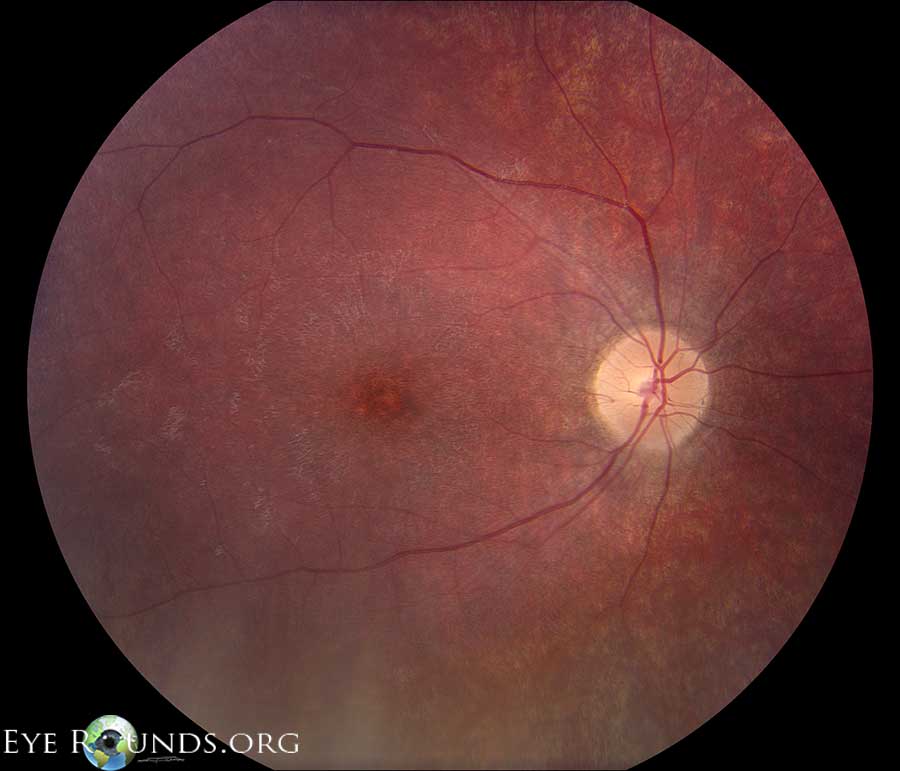
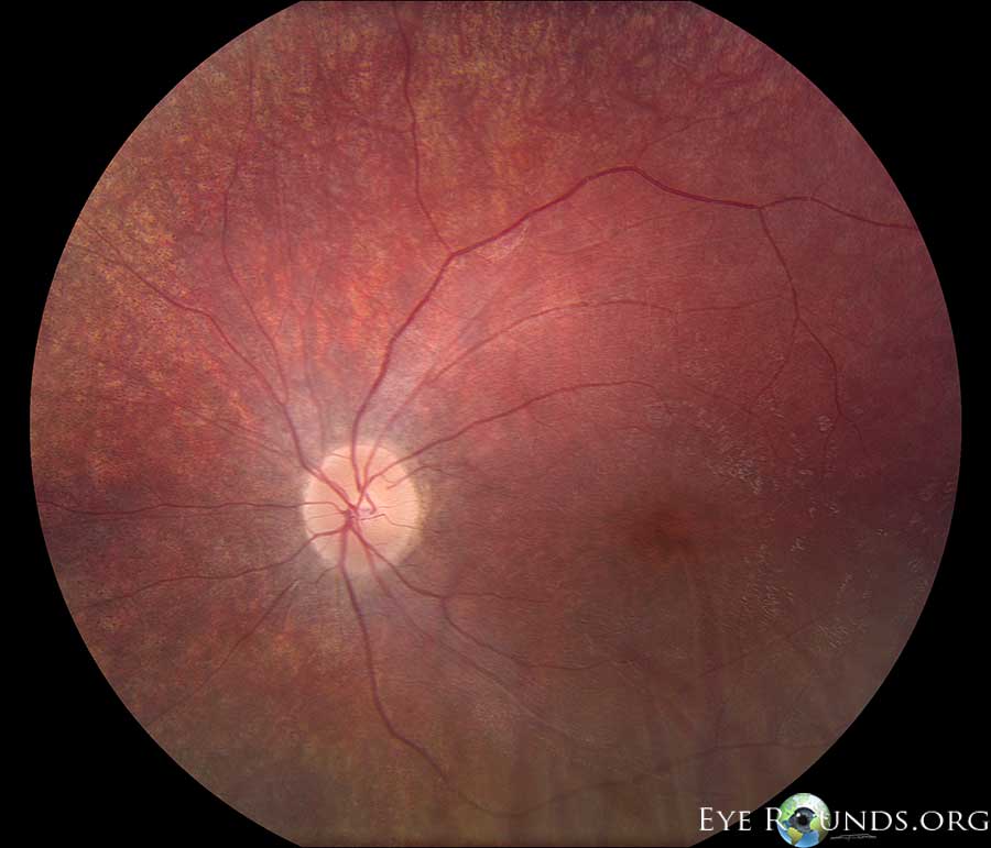
7-year-old female with molecularly confirmed CLN3-associated Batten Disease.

Fundus photos demonstrate mild optic nerve pallor, central macular mottling, attenuated arterioles and peripheral retinal granularity. There was 1+ vitreous cell present. Vision was 20/800 in the right eye and hand motion in the left. Batten disease is a fatal autosomal recessive neurodegenerative disease and is the most common neuronal ceroid lipofuscinosis.
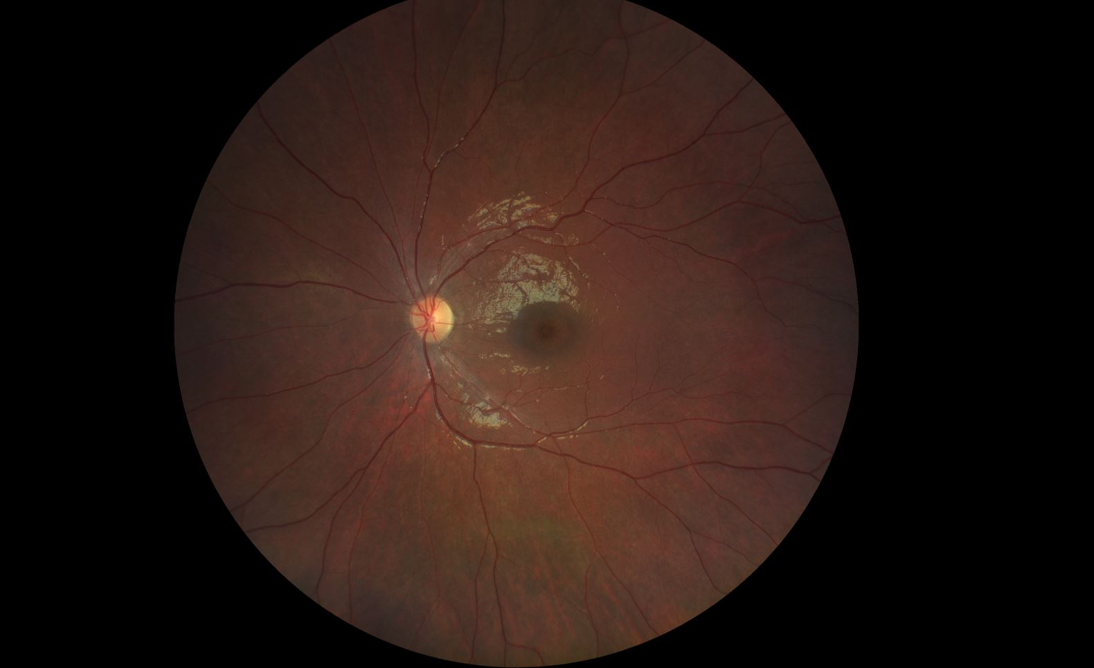
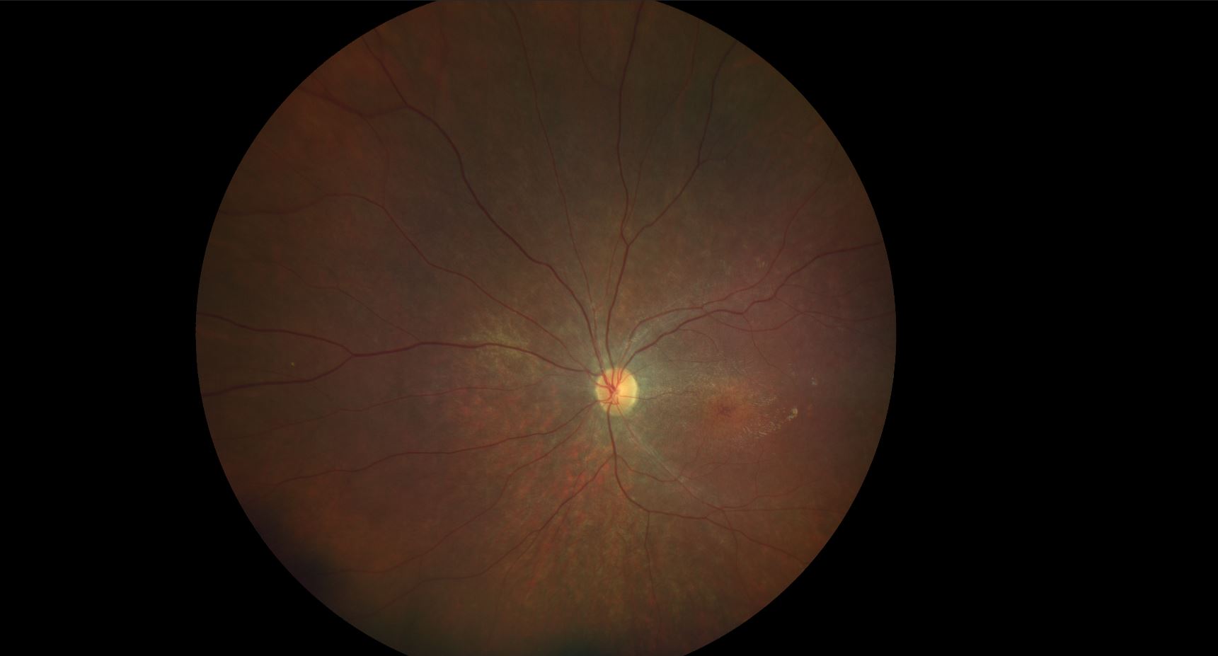
OMIM #204200


Bullseye maculopathy in Batten Disease

Bullseye maculopathy in Batten Disease

Category(ies): Inherited Eye Diseases, Retina / Vitreous
Contributor: Ella J. Gehrke, MD; Arlene V. Drack, MD
Photographer: Douglas B. Critser, CRA, OCT-C, FOPS
Posted: 02/19/2025

Fundus photo demonstrates flat, dark pink disc with bullseye maculopathy. Vision at time of diagnosis was 20/40 OU BCVA in a 9-year-old female. Bullseye maculopathy associated with Batten disease at time of first presentation.

Fundus photo demonstrates flat, dark pink disc with bullseye maculopathy. Vision at time of diagnosis was 20/40 OU BCVA in a 9-year-old female.
The second photo taken from the same child, 5 years after the first photo. In this second photo, the macula has a more degenerative hyperpigmented appearance with epiretinal membrane and a loss of the
The second photo taken from the same child, 5 years after the first photo. In this second photo, the macula has a more degenerative hyperpigmented appearance with epiretinal membrane and a loss of the "bullseye" appearance. This emphasizes that the maculopathy in the first photo is not diagnostic and may not be present in every case. There is also more nerve pallor.