This site uses tracking information. Visit our privacy policy. Click to agree to this policy and not see this again.

Ophthalmology and Visual Sciences

Cilioretinal Artery Occlusion

Cilioretinal Artery Occlusion

Category(ies): Retina/Vitreous
Contributor: Austin Fox, MD and Stephen Russell, MD
Photographer: Susan Wright

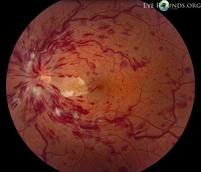
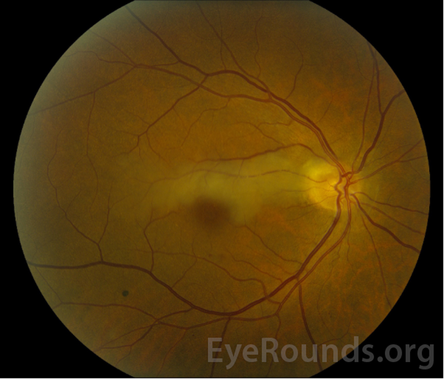
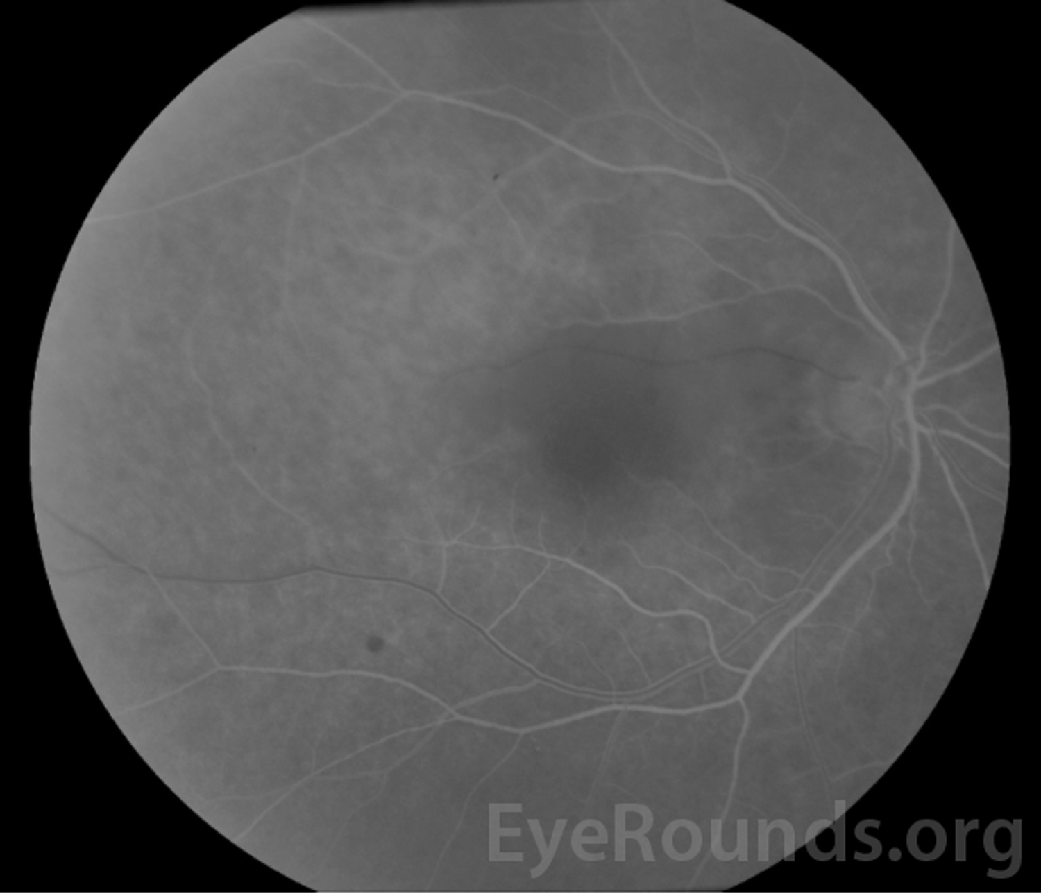
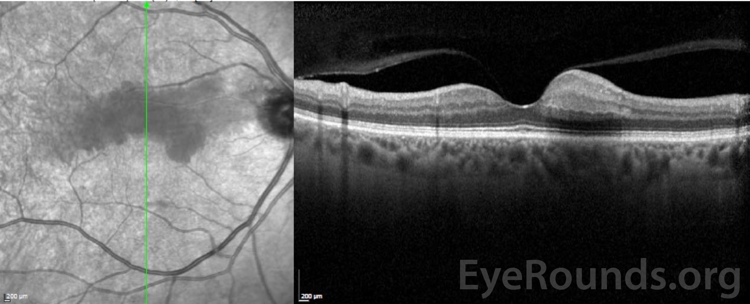
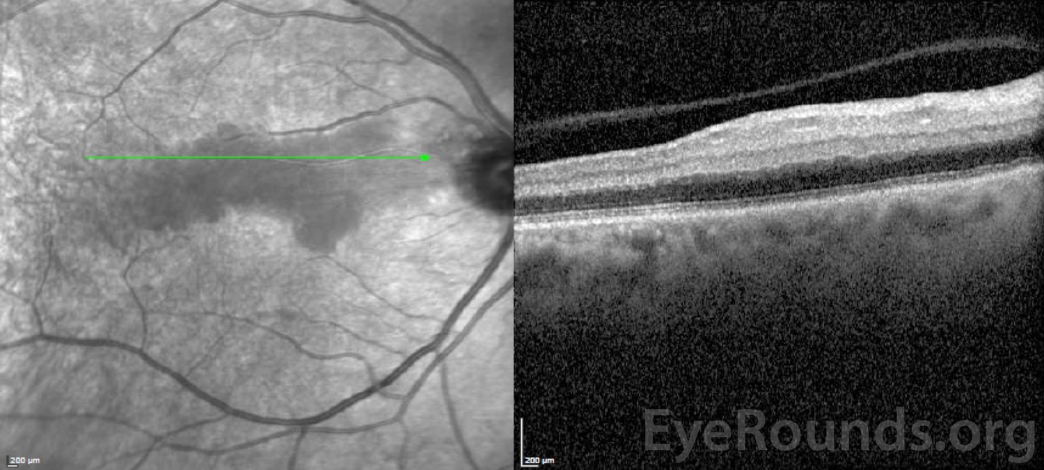
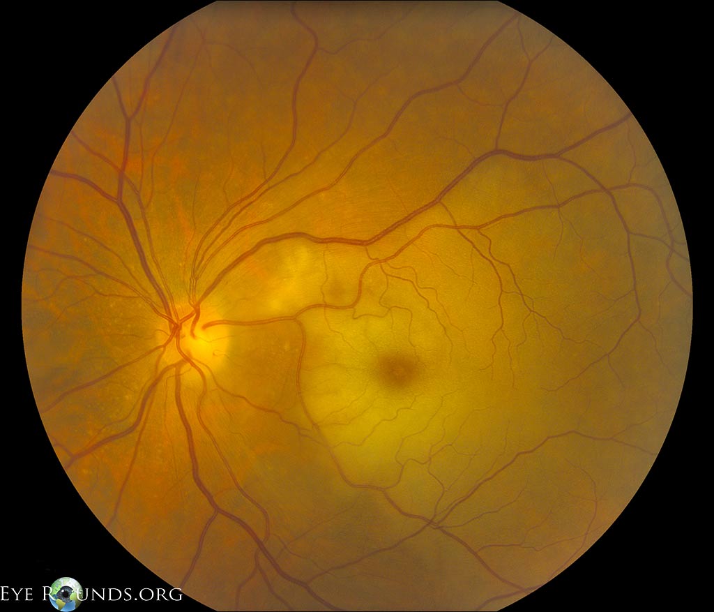
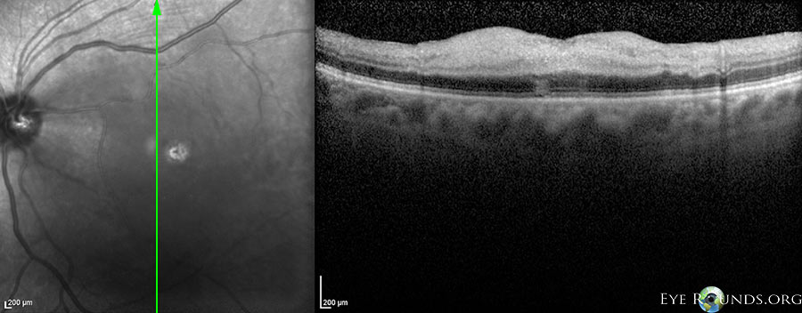
A 67-year-old man with history of type 2 diabetes mellitus, hyperlipidemia, and uncontrolled hypertension presented with a central visual field deficit of sudden onset in his right eye. Visual acuity on presentation was 20/25 in both eyes, and funduscopic examination revealed an isolated cilioretinal artery occlusion (CLRAO) in his right eye (Figure 1). Work-up revealed significant right internal carotid artery stenosis (>50%). A right carotid endarterectomy was performed, and vascular risk factors were optimized. Visual acuity was stable at 20/25 one month after presentation.

CLRAO accounts for ~5-7% of retinal artery occlusions and is usually associated with a central retinal vein occlusion (CRVO) or anterior ischemic optic neuropathy (AION). [1-3] Less commonly, CLRAOs may occur as an isolated phenomenon as in this case. Isolated CLRAOs typically have a better visual prognosis than those associated with a CRVO or AION with 90% of patients achieving a visual acuity of 20/40 or better. [3]

Multiple small emboli are seen within the cilioretinal artery
Figure 1. Retinal whitening extends from the superotemporal optic nerve through the macula superior to the fovea. Multiple small emboli are seen within the cilioretinal artery.
 Fluorescein angiography of the right eye shows a filling defect of the cilioretinal artery superior to the fovea
Figure 2. Fluorescein angiography of the right eye shows a filling defect of the cilioretinal artery superior to the fovea.
 Optical coherence tomography shows retinal edema of the inner retinal layers superior to the fovea in the distribution
Figure 3. Optical coherence tomography shows retinal edema of the inner retinal layers superior to the fovea in the distribution of the cilioretinal artery.
 Optical coherence tomography shows retinal edema of the inner retinal layers superior to the fovea in the distribution
Figure 3. Optical coherence tomography shows retinal edema of the inner retinal layers superior to the fovea in the distribution of the cilioretinal artery.
 Optical coherence tomography shows retinal edema of the inner retinal layers superior to the fovea in the distribution
Contributor: Jesse Vislisel, MD, The University of Iowa
71-year-old woman presenting with a cilioretinal artery occlusion. Note the retinal whitening secondary to inner retinal edema in the distribution of cilioretinal artery perfusion. There is a prominent cherry-red spot due to intact choroidal circulation.
 Optical coherence tomography shows retinal edema of the inner retinal layers superior to the fovea in the distribution
Contributor: Jesse Vislisel, MD, The University of Iowa
71-year-old woman presenting with a cilioretinal artery occlusion. On OCT, there is inner retinal thickening and blurring of retinal layers due to edema.
Reference:
  • Brown GC, Shields JA. Cilioretinal arteries and retinal arterial occlusion. Arch Ophthalmol. 1979;97:84–92.
  • Hayreh SS. Pathogenesis of occlusion of the central retinal vessels. Am J Ophthalmol 1971;72:998–1011
  • Brown GC, Moffat K, Cruess A, Magargal LE, Goldberg RE. Cilioretinal artery obstruction. Retina. 1983;3(3):182-7

Image Permissions:
Creative Commons

Ophthalmic Atlas Images by EyeRounds.org, The University of Iowa are licensed under a Creative Commons Attribution-NonCommercial-NoDerivs 3.0 Unported License.