

Episcleritis
Contributor: William Charles Caccamise, Sr, MD, Retired Clinical Assistant Professor of Ophthalmology, University of Rochester School of Medicine and Dentistry
*Dr. Caccamise has very generously shared his images of patients taken while operating during the "eye season" in rural India as well as those from his private practice during the 1960's and 1970's. Many of his images are significant for their historical perspective and for techniques and conditions seen in settings in undeveloped areas.
Category: External Disease
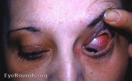
Acute episcleritis - watch for development of nodular scleritis.
Episcleritis with keratitis

This patient is typical of the majority of episcleritis cases, i.e. a female in her late 40s. Corneal infiltration can be seen in juxtaposition to the episcleritis.
Bilateral episcleritis - with ulcerative colitis
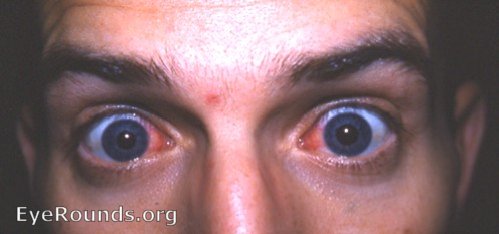
This young scholar had recurrent episcleritis - bilateral - with flare-ups in his ulcerative colitis. Googling " ulcerative colitis and episcleritis " will produce several pertinent articles.
Episcleritis, sectoral
Contributor: Hilary A. Beaver, MD and Jordan M. Graff, MD, University of Iowa
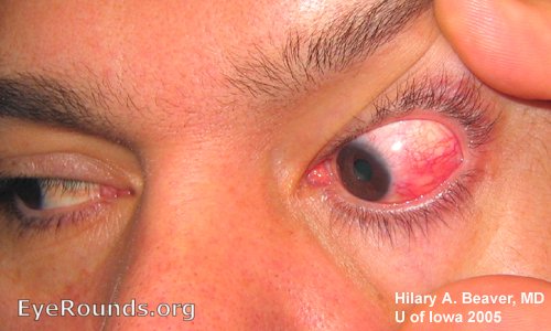
There is a localized episcleritis, without nodularity. The patient has no known systemic disease (including no inflammatory bowel disease nor rheumatoid arthritis). Note the violacious color of the inflammed section. The remainder of the sclera and episclera in this eye and the contralateral eye remain white and quiet.

Ophthalmic Atlas Images by EyeRounds.org, The University of Iowa are licensed under a Creative Commons Attribution-NonCommercial-NoDerivs 3.0 Unported License.