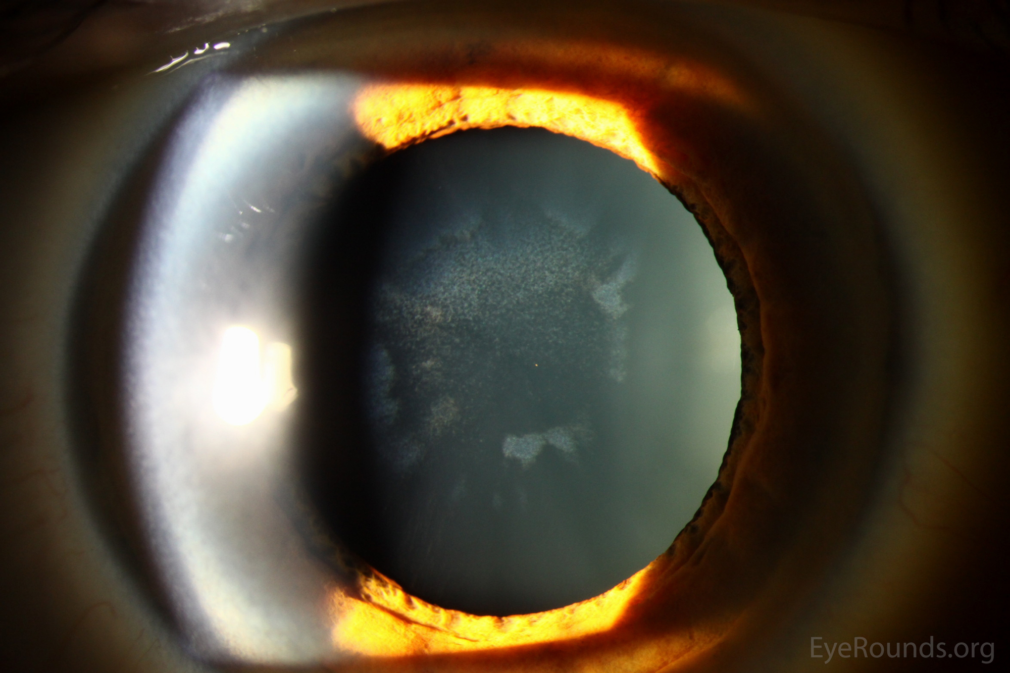
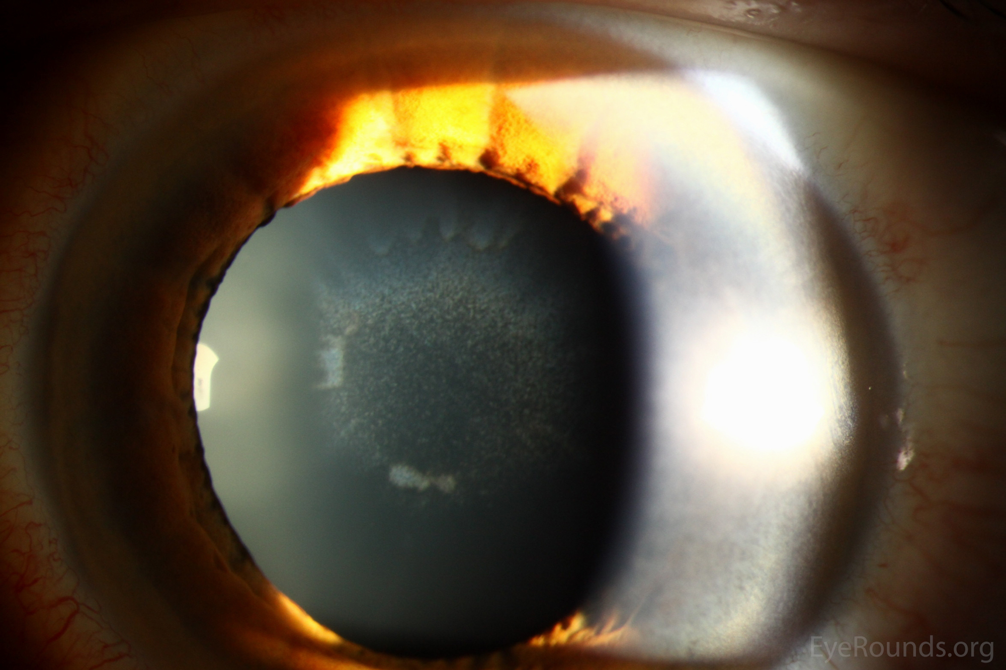
Figure 1a. Slit lamp photographs of sunflower cataracts of the right eye. Note the ring-shaped, anterior capsular opacities that resemble a "sunflower," a finding that is classically associated with Wilson's Disease.
Figure 1b. Slit lamp photographs of sunflower cataracts of the left eye. Note the ring-shaped, anterior capsular opacities that resemble a "sunflower," a finding that is classically associated with Wilson's Disease.
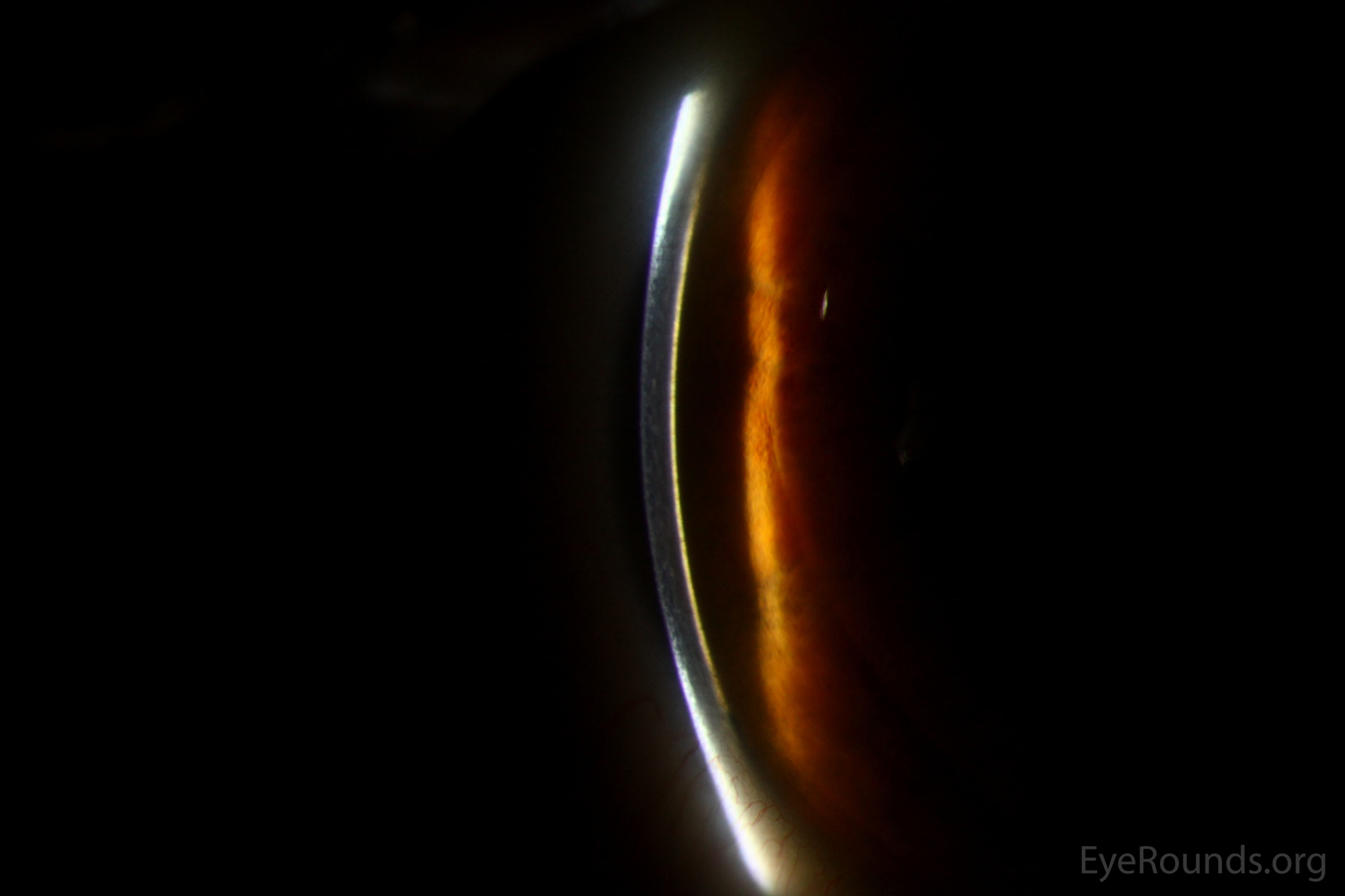
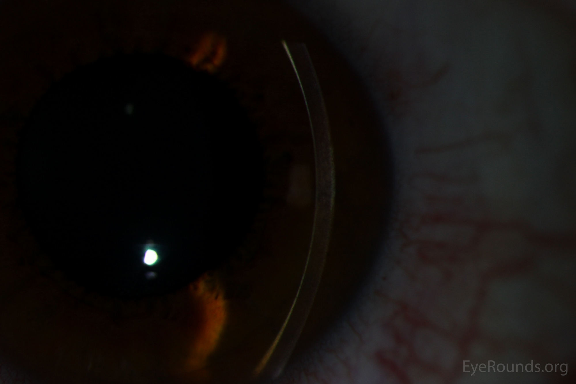
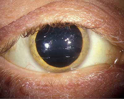
Figure 2a. Slit lamp photographs of a Kayser-Fleischer ring. Note the prominent golden/copper hue of the peripheral corneal endothelium—a result of copper deposition, primarily in Descemet membrane.
Figure 2b. Slit lamp photographs of a Kayser-Fleischer ring. Note the prominent golden/copper hue of the peripheral corneal endothelium—a result of copper deposition, primarily in Descemet membrane.
Figure 2c. Note how the posterior corneal reflex loses the prominent golden/copper hue centrally but maintains the dull metallic sheen superiorly and inferiorly (seen most prominently inferiorly in this photograph)
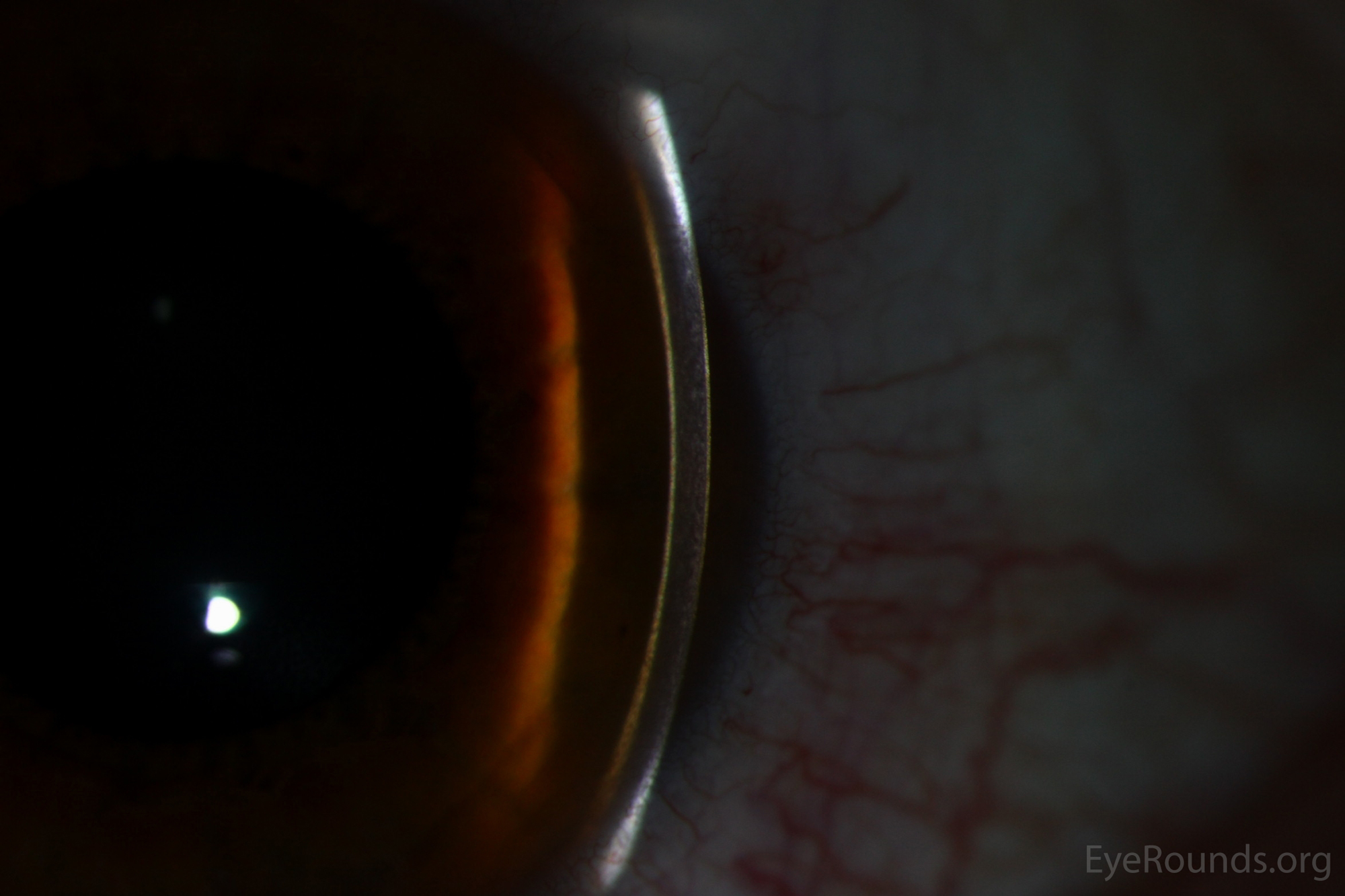
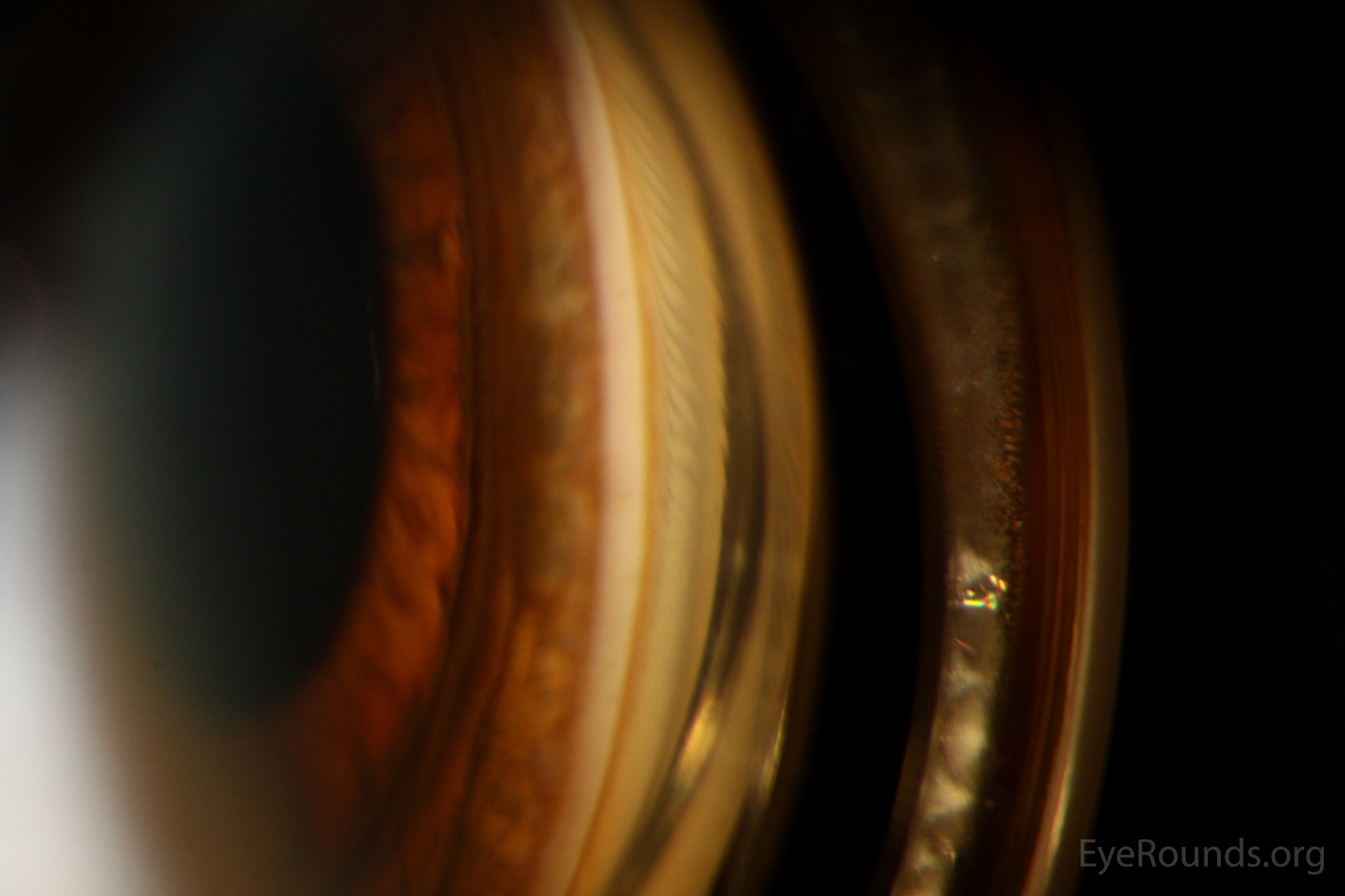
Figure 3. Slit lamp gonioscopy photographs of a Kayser-Fleischer ring. Note the dark brown/gold/copper band located anterior to the trabecular meshwork in the peripheral cornea.
University of Iowa
Roy J. and Lucille A. Carver College of Medicine
Department of Ophthalmology and Visual Sciences
200 Hawkins Drive
Iowa City, IA 52242
University of Iowa
Roy J. and Lucille A. Carver College of Medicine
Department of Ophthalmology and Visual Sciences
200 Hawkins Drive
Iowa City, IA 52242