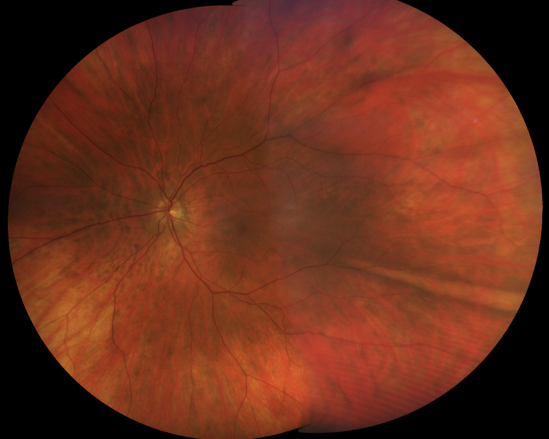
The long ciliary nerves carry somatosensory fibers from the cornea, iris, and ciliary body that contribute to the nasociliary nerve (branch of V1). They also carry post-ganglionic sympathetic fibers from the superior cervical ganglion which provide sympathetic innervation to the dilator pupillae muscle. They pierce the sclera laterally or inferolaterally to the optic nerve and course anteriorly between the sclera and choroid. They are sometimes appreciated near the 3 and 9 o’clock positions on dilated fundus exam.

Ophthalmic Atlas Images by EyeRounds.org, The University of Iowa are licensed under a Creative Commons Attribution-NonCommercial-NoDerivs 3.0 Unported License.


Address
University of IowaLegal
Related Links