This site uses tracking information. Visit our privacy policy. Click to agree to this policy and not see this again.

Ophthalmology and Visual Sciences

Marshall Syndrome

Marshall Syndrome

Category(ies): Retina, Glaucoma
Contributor: Brittni A. Scruggs, MD, PhD and John H. Fingert, MD, PhD

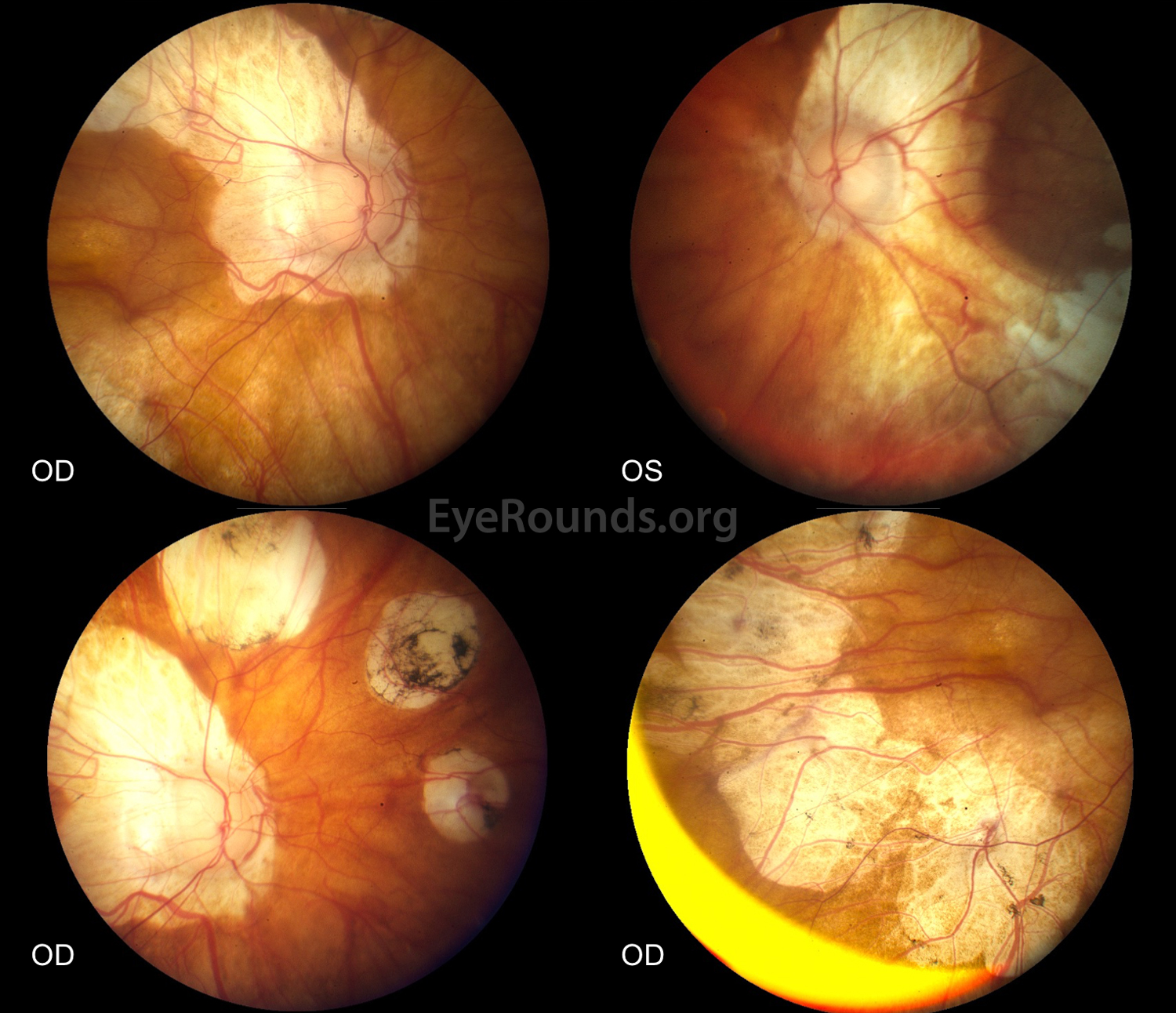
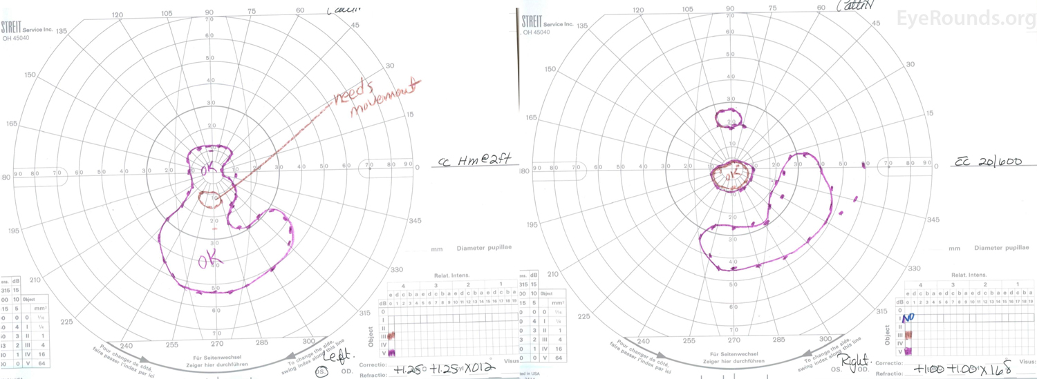
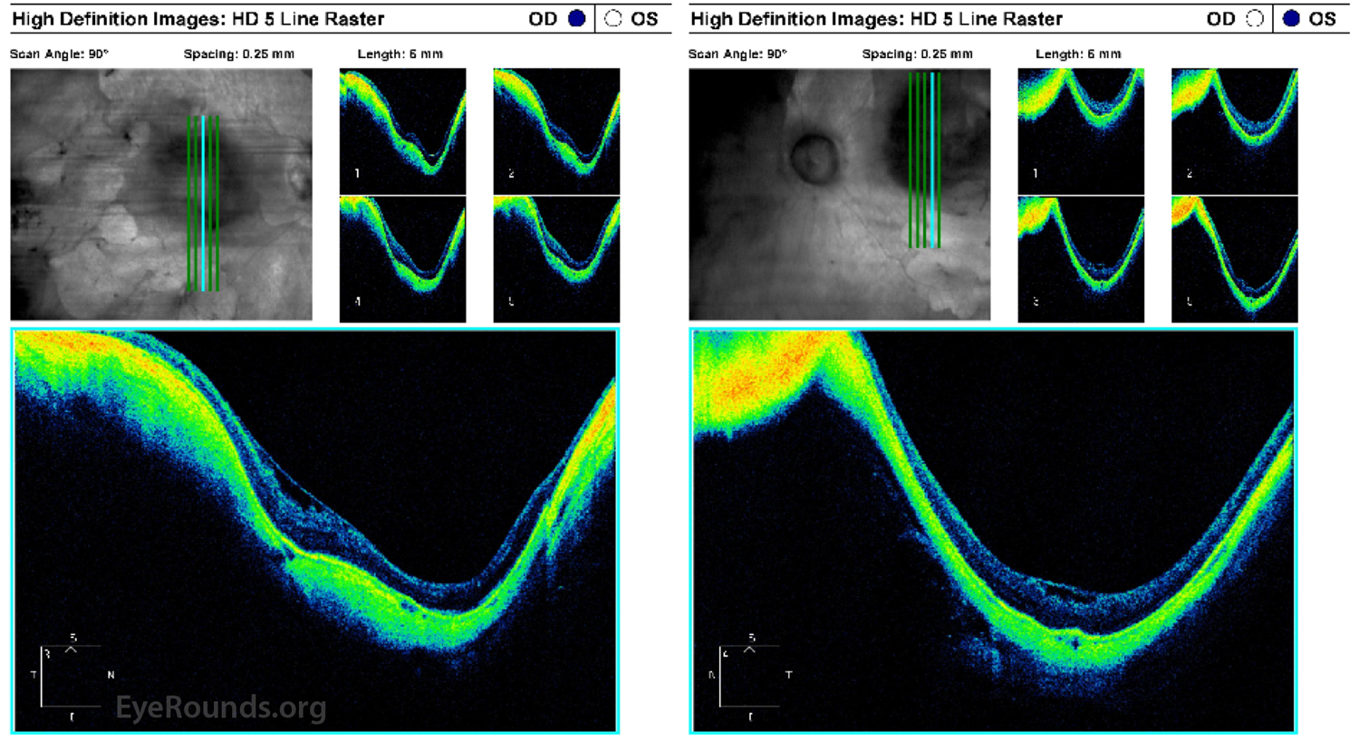
This patient had a lifelong history of profound hearing loss and vision loss in the left eye (OS) greater than right eye (OD) in the setting of Marshall syndrome with high myopia and aphakic glaucoma in both eyes (OU). She underwent strabismus surgery for esotropia as a child and bilateral extracapsular cataract extraction when she was 27-years-old. Her visual acuity progressively decreased to 20/600 OD and hand motions OS due to myopic degeneration OU. Dilated funduscopic examination showed numerous, scattered chorioretinal atrophic regions, vascular attenuation, and large posterior staphylomas OU (Figure 1). Goldmann visual fields showed a small, preserved central island of vision OU (Figure 2). Optical coherence tomography of the macula showed staphylomatous changes with severe retinal and choroidal thinning OU (Figure 3).

Marshall syndrome is a rare autosomal dominant syndrome that presents with pathologic myopia, congenital cataracts, sensorineural hearing loss, hypertelorism, and a flattened nasal bridge. Patients with Marshall syndrome have COL11A1 gene mutations leading to abnormalities in the production of collagen; mutations in this gene are also found in Type 2 Stickler's syndrome. Strabismus, retinal detachments, arthritis, and ectodermal abnormalities are common in Marshall syndrome [1].

4 fundus photos showing marshall syndrome
Figure 1. Color fundus photography, OU.There are large areas of choroidal atrophy OU, including atrophy along the superior and inferior arcades OU and atrophic lesions encircling the macula OU. The patient has significant posterior staphylomas OS>OD. There is marked retinal vascular attenuation. The optic nerves are tilted and have diffuse, moderate pallor OU.
 Goldmann kinetic perimetry
Figure 2. Goldmann kinetic perimetry, OU.OD (left image): There is a central island of preserved vision to the V4e and III4e isopters with a temporal island present only to the V4e isopter. OS (right image): There is a central and inferior island of preserved vision to the V4e isopter.These profound visual fields defects are due to the patient's pathogenic myopia and severe aphakic glaucoma OU.
OCT
Figure 3. Optical coherence tomography of the macula, OU. Posterior staphylomas are present OU, and the retina is attached OU. There is no intraretinal or subretinal fluid OU. There is marked thinning of the choroid and retina OU, especially in regions corresponding to atrophy along the inferior arcades OU. Mild epiretinal membranes are present centrally OU.
References:
  1. Khalifa O, Imtiaz F, Ramzan K, Allam R, Hemidan AA, Faqeih E, et al. Marshall syndrome: further evidence of a distinct phenotypic entity and report of new findings. Am J Med Genet A. 2014;164A(10):2601-6.

Image Permissions:
Creative Commons

Ophthalmic Atlas Images by EyeRounds.org, The University of Iowa are licensed under a Creative Commons Attribution-NonCommercial-NoDerivs 3.0 Unported License.