This site uses tracking information. Visit our privacy policy. Click to agree to this policy and not see this again.

Ophthalmology and Visual Sciences

Sectoral retinitis pigmentosa

Sectoral retinitis pigmentosa

Category(ies): Retina, Inherited Eye Disease
Contributor: Jesse Vislisel, MD
Photographer: Brice Critser, CRA

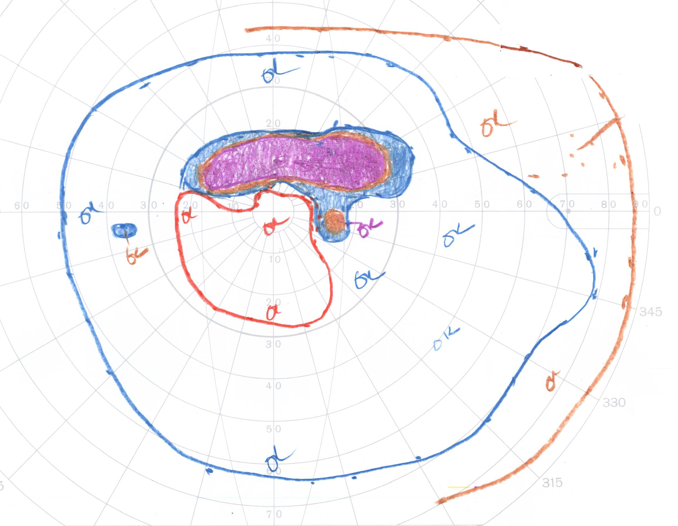
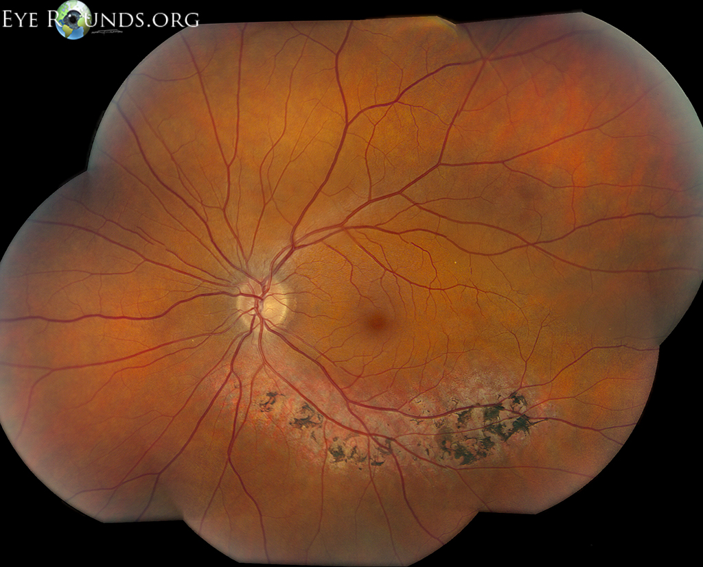
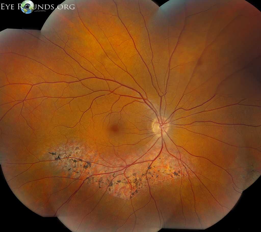
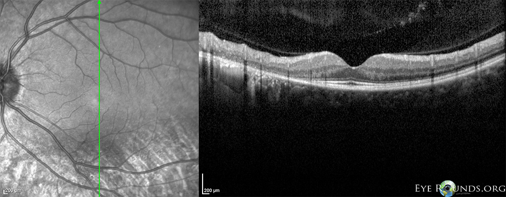
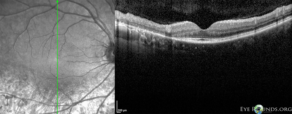
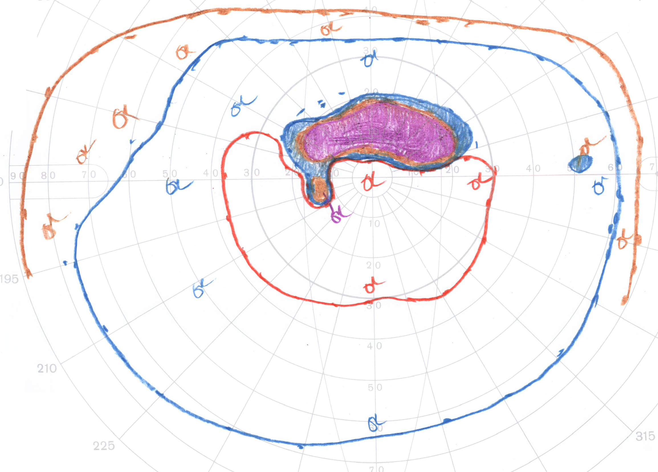
Sectoral retinitis pigmentosa (RP) is a variant of RP in which there is a regional distribution of the retinal degeneration. It can be differentiated from acquired pigmentation (due to trauma, inflammation, or vascular insult) by its symmetry between the eyes. These photos depict segmental RP in a patient with autosomal dominant RP secondary to a rhodopsin defect. Note the prominent areas of chorioretinal atrophy with bone-spicule-like pigmentation along the inferotemporal arcade. The Goldmann visual fields show superior scotomata that correspond to this area of degeneration. The OCT shows marked outer retinal and retinal pigmented epithelial (RPE) thinning in the affected area.

Sectoral retinitis pigmentosa (RP) montage os
OS - These photos depict segmental RP in a patient with autosomal dominant RP secondary to a rhodopsin defect. Note the prominent areas of chorioretinal atrophy with bone-spicule-like pigmentation along the inferotemporal arcade.
Sectoral retinitis pigmentosa (RP) montage od
OD - These photos depict segmental RP in a patient with autosomal dominant RP secondary to a rhodopsin defect. Note the prominent areas of chorioretinal atrophy with bone-spicule-like pigmentation along the inferotemporal arcade.
Sectoral retinitis pigmentosa (RP) OCT os
OS - The OCT shows marked outer retinal and retinal pigmented epithelial (RPE) thinning in the affected area.
Sectoral retinitis pigmentosa (RP) OCT OD
OD - The OCT shows marked outer retinal and retinal pigmented epithelial (RPE) thinning in the affected area.
Sectoral retinitis pigmentosa (RP) Goldmann  os
OS - The Goldmann visual fields show superior scotomata that correspond to this area of degeneration. The OCT shows marked outer retinal and retinal pigmented epithelial (RPE) thinning in the affected area..
Sectoral retinitis pigmentosa (RP) Goldmann  od
OD - The Goldmann visual fields show superior scotomata that correspond to this area of degeneration. The OCT shows marked outer retinal and retinal pigmented epithelial (RPE) thinning in the affected area..