
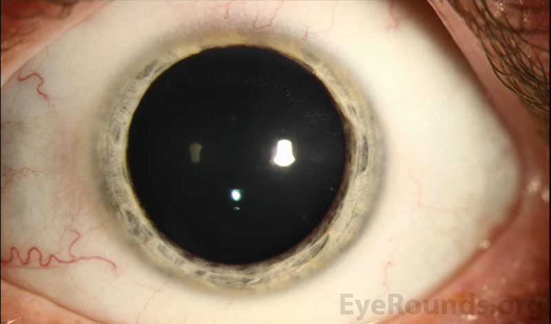
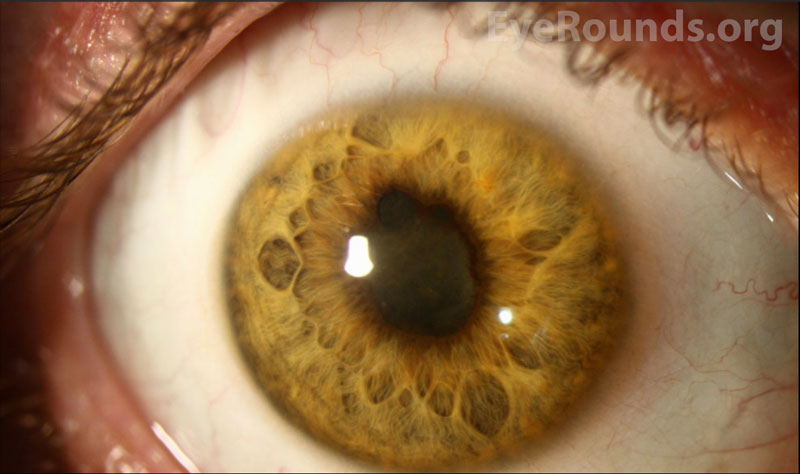
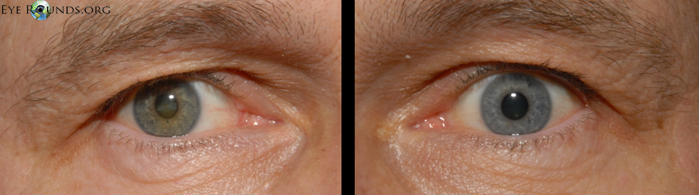
These external photographs demonstrate an acquired hyperchromic heterochromia due to a traumatic intraocular iron foreign body in the right eye. Chronic deposition of iron within the iris stroma resulted in the gradual darkening of the native light-colored iris.
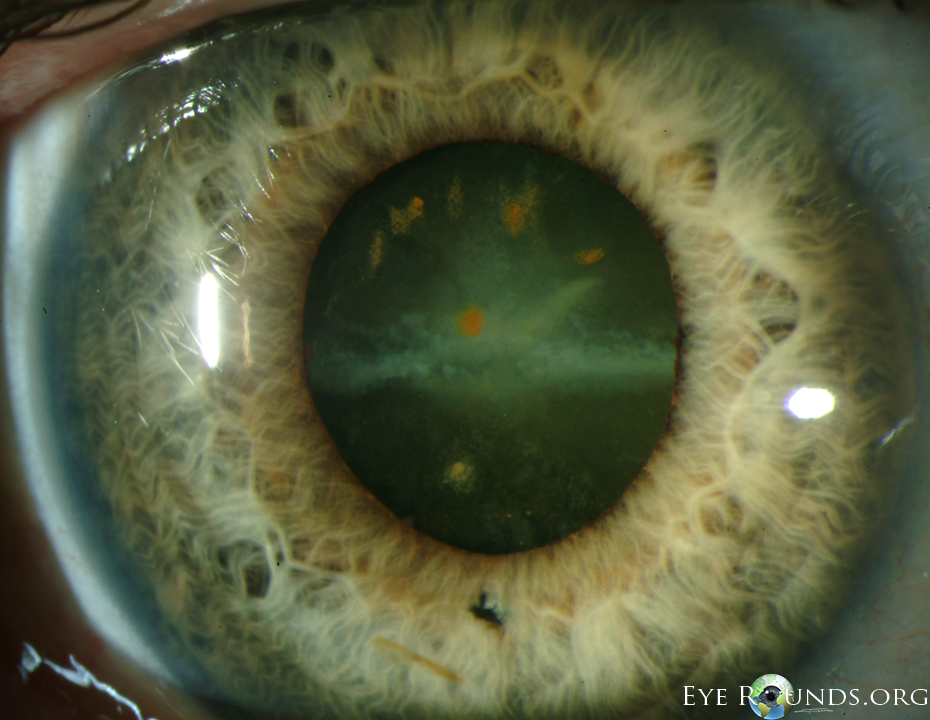
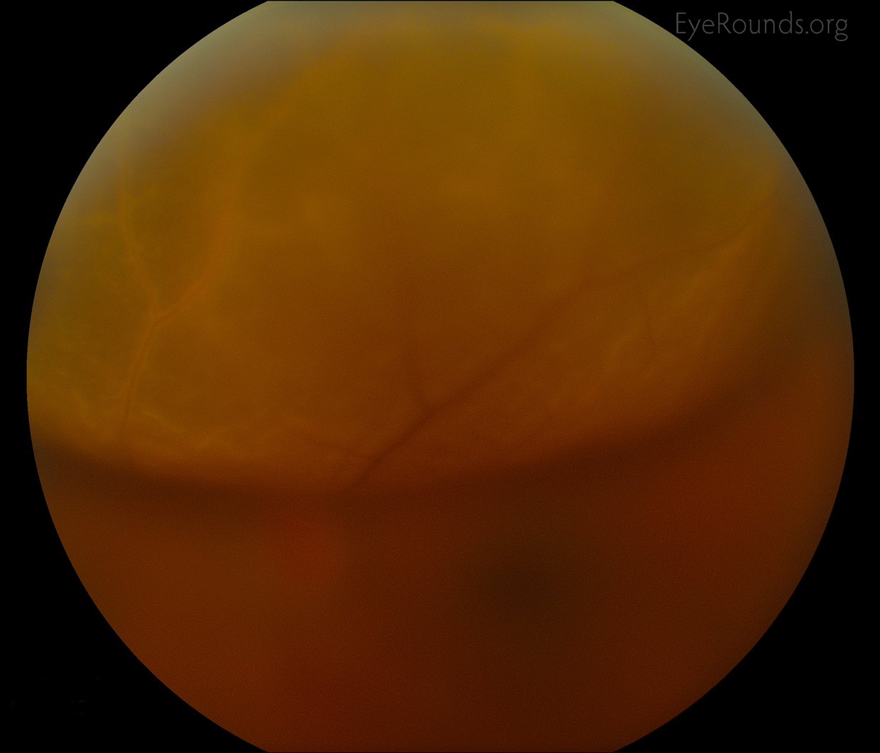
Siderosis bulbi is caused by retention and oxidation of an iron-containing intraocular foreign body. Clinical features include cataract, rust-colored anterior subcapsular deposits, iris heterochromia (affected side is darker), pupillary mydriasis, and depressed electroretinogram (ERG) amplitudes. Other potential sequelae include pigmentary retinopathy, retinal microangiopathy, and open-angle glaucoma. If the cataract and foreign body are removed and there is no macular trauma, the visual prognosis is excellent.
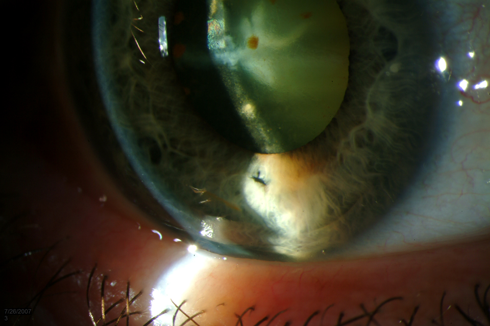
These photos demonstrate iris heterochromia and rust-colored anterior subcapsular deposits in a patient with a history of metal-on-metal ocular trauma. An iris defect and a full-thickness corneal wound are also present inferiorly.

Ophthalmic Atlas Images by EyeRounds.org, The University of Iowa are licensed under a Creative Commons Attribution-NonCommercial-NoDerivs 3.0 Unported License.

Address
University of IowaLegal
Related Links
