Inability to obtain 20/20 vision with refraction in the left eye
This 11-year-old female presented as a referral from her local optometrist for inability to improve the vision in the left eye beyond 20/30 with refraction despite an otherwise normal eye exam. The patient was asymptomatic and felt that her vision was the same as it had always been. She had never had an eye exam before seeing her local optometrist and presented there for a routine exam to establish care without specific ocular complaints.
Monofixation syndrome and mild anisometropic amblyopia OS
Given that the patient was asymptomatic from her condition and only a mild, subjectively insignificant improvement in visual acuity in the left eye could be obtained with cycloplegic refraction, glasses were not prescribed. The effect of glasses to treat the decreased vision on the left was subjectively insignificant because she had foveal suppression in this eye under binocular conditions related to monofixation syndrome. Her amblyopia in the left eye was very mild and she was beyond the age range where occlusion or penalization therapy would be considered given her visual maturity.
Monofixation syndrome is an adaptive sensory state that occurs secondary to disruption of binocularity during development of the visual system (usually within the first eight to ten years of life). It is characterized by central foveal suppression in one eye with maintained binocular fusion of the peripheral visual fields. It is caused by small-angle strabismus (< 10 PD) or a mild to moderate degree of unilateral retinal image blur (i.e., anisometropia). The central macula and fovea have small receptive fields and potential for high spatial resolution that make it prone to perceive differing interocular visual inputs from relatively small differences in image clarity or retinal image position between the two eyes. This leads to monocular central suppression in monofixation syndrome. In contrast, the more peripheral macula and retina have larger receptive fields and lower spatial resolution that allows for larger degrees of retinal image discrepancy while still being able to maintain binocular fusion. This is the mechanism by which peripheral fusion is maintained in monofixation syndrome. The size of the central suppression scotoma is directly proportional to the degree of interocular image disparity. If there is severe unilateral image blur (i.e., dense cataract) or large-angle strabismus that exceeds the amount of image disparity that allows for fusion in the peripheral visual fields, suppression of visual input from the entire eye may occur and amblyopia may develop.
Monofixation syndrome is typically encountered in patients with small-angle strabismus (<10 PD), anisometropic amblyopia, unilateral astigmatism (meridional anisometropia) or unilateral partial or mild media opacity (i.e., cataract). These patients have motor fusion, but will often have a relatively large underlying phoria in addition to the small tropia that is frequently present. Stereo acuity is typically maintained in the range of 3000-700 arc seconds. The unilateral central suppression scotoma is usually between 2-5 degrees of visual field. Of note, however, this is a facultative scotoma. It is only present under binocular conditions and as soon as the dominant, fixating eye is occluded, the central scotoma of the non-dominant eye vanishes. Patients with monofixation syndrome typically have some degree of amblyopia that can range from mild to severe.
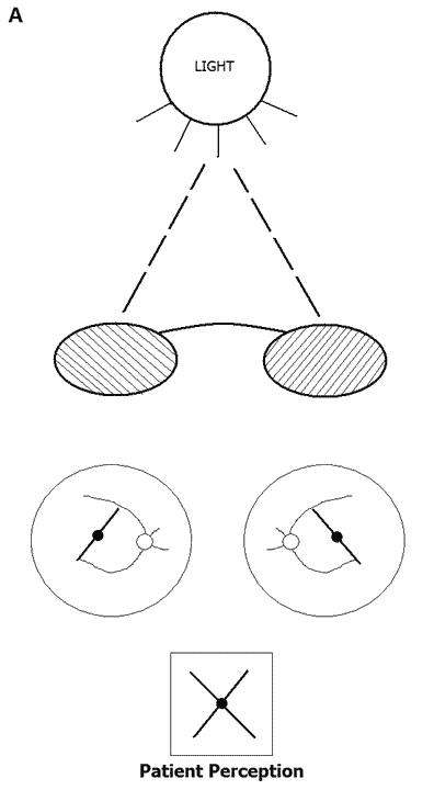
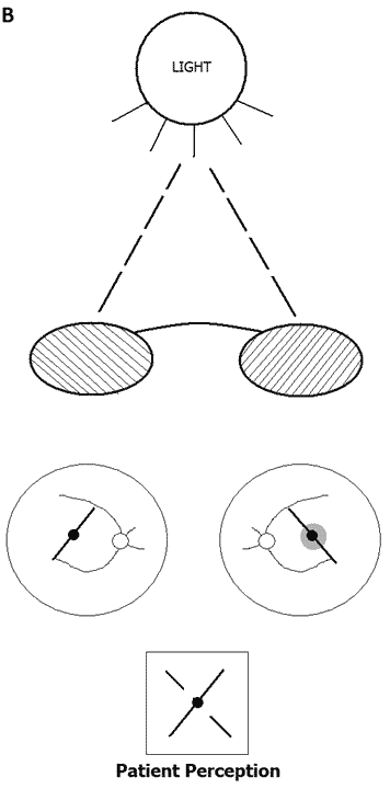
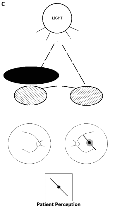
Bagolini lenses (Figure 1): Bagolini striated lenses are a sensory test that present linear streaks of light to each eye that are oriented 90 degrees apart with a central fixation light that is transected by each streak. In normal binocular vision, the patient will describe the central fixation light that is crossed by each streak. In monofixation syndrome, the patient will describe the central fixation light that is crossed by only one streak from the dominant fixating eye and will perceive the other streak from the non-dominant eye to have a gap where it would cross the fixation light that represents the suppression scotoma. This gap would disappear if the dominant eye were occluded demonstrating the facultative nature of the suppression scotoma.
Figure 1[4]
Bagolini striated lenses convert a fixation light into a streak of light oriented perpendicular to the striations on the lenses. Pictured here, the striations over OD are oriented at 135 degrees creating a streak of light oriented at 45 degrees as perceived by the patient and the striations over OS are oriented at 45 degrees creating a streak of light oriented at 135 degrees as perceived by the patient. In the absence of pathology, the patient will perceive a cross of light through the center of the fixation light. |
In a patient with monofixation syndrome, the fixating eye (pictured as OD) will perceive a continuous streak of light intersecting the fixation light and the non-fixating eye (pictured as OS) will perceive the streak of light as having a gap in it around fixation that represents the central suppression scotoma in this eye. |
Given that the suppression scotoma in the non-fixating eye (OS) is facultative, covering the fixating eye (OD) will result in resolution of the scotoma and the patient will perceive a continuous streak of light intersecting the fixation light in this eye. |
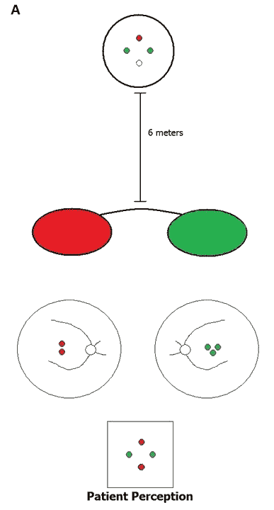
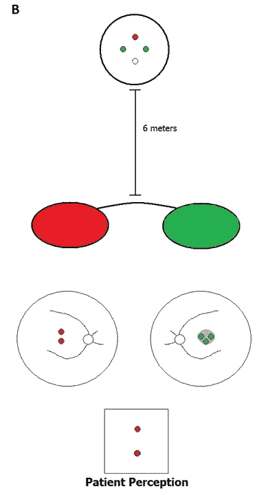
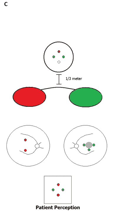
Worth 4-dot (Figure 2): Worth 4-dot is another sensory test that is useful in evaluating monofixation syndrome. When tested at distance (6 meters), the dots are 1.25 degrees apart. When tested at near (1/3 meter), the dots are 6 degrees apart. The central foveal suppression scotoma in monofixation syndrome is 5 degrees or less. When shown the Worth 4-dot at distance, all of the dots will fall within this scotoma and it will appear as if the patient is suppressing one eye. However, when shown the Worth 4-dot at near, the dots are 6 degrees apart and will fall outside of the central suppression scotoma and the patient will appear to have binocular fusion. This demonstrates unilateral central foveal suppression with binocular peripheral fusion.
Figure 2[4]
The Worth 4-dot consists of two green lights, one red light, and one white light as seen above. The patient wears red/green glasses, typically with the red lens OD and the green lens OS (pictured). The red light is seen by the eye with the red filter (OD), the green lights are seen by the eye with the green filter (OS) and the white light is seen by both eyes and can be perceived as a combination of red and green, can alternate between red and green or can be seen as the color filter over the dominant/fixating eye (pictured in figure as OD). Figure 2A is a normal Worth 4-dot with OD as the dominant/fixating eye. |
Worth 4-dot tested at a distance of 6 meters will separate the dots by 1.25 degrees. In monofixation syndrome, the central suppression scotoma (pictured OS) occupies the central 2-5 degrees which places the dots perceived by this eye within it and it appears as if the patient is suppressing this eye. |
|
Cover testing: In patients too young to perform sensory testing, monofixation syndrome can be suspected if there is a small tropia of less than 10 PD elicited by cover-uncover testing that builds to a larger angle phoria as they are dissociated and broken-down with alternate cover testing to show the full deviation. The presence of a phoria is an indication of some form of binocular fusion. Monofixation syndrome has been called phoria-tropia syndrome. The small-angle tropia can be difficult to pick up on testing.
4 prism diopter base-out test: The 4 prism diopter base-out test is another diagnostic technique that can be used to evaluate for a central suppression scotoma. To perform this test, a 4 PD base-out prism is placed in front of one eye and then the other under binocular conditions and ocular motor responses are observed. In a normal patient, a version (bilateral) movement of both eyes away from the eye covered by the prism followed by a unilateral fusional convergence movement (adduction) of the eye not covered by the prism will be observed. This response is the same regardless of the eye covered. In monofixation syndrome, if the prism is placed in front of the non-fixating eye, no ocular motor response is observed as the displaced image falling on the retina of this eye has not been moved outside of the suppression scotoma. If the prism is placed in front of the fixating eye, a refixation version will be observed, but there will be no fusional convergence movement of the contralateral eye.
The treatment for monofixation syndrome is the treatment of the amblyopia associated with it. Correction of refractive error or amelioration of media opacities can be addressed. If significant amblyopia is present and the patient is of appropriate age, occlusion or penalization therapy may be indicated. Monofixation syndrome is considered to be a positive surgical outcome of congenital heterotropias given that this sensory state that has some degree of binocular fusion (even though it is not bifoveal) making the surgical outcome more stable and the residual small-angle heterotropia is cosmetically favorable. Strabismus surgery for the small-angle heterotropia seen in monofixation syndrome is not indicated.
Etiology
|
Signs
|
Symptoms
|
Treatment
|
Strabismus
Anisometropia
Meridional anisometropia
Stimulus deprivation (e.g., cataract)
Amblyopia
Kirkpatrick CA, Scott WE. Monofixation Syndrome: 11-year-old female referred after inability to improve vision with refraction in the left eye. Feb 25, 2015; Available from: https://eyerounds.org/cases/205-monofixation-Syndrome.htm

Ophthalmic Atlas Images by EyeRounds.org, The University of Iowa are licensed under a Creative Commons Attribution-NonCommercial-NoDerivs 3.0 Unported License.

Address
University of IowaLegal
Related Links