This site uses tracking information. Visit our privacy policy. Click to agree to this policy and not see this again.

Ophthalmology and Visual Sciences

Chronic Ocular Graft Versus Host Disease

Chronic Ocular Graft Versus Host Disease

Andrew S. Goldstein, BS; Caroline W. Wilson, MD; Mark A. Greiner, MD

INITIAL PRESENTATION

Chief Complaint

Bilateral eye irritation, redness, and light sensitivity

History of Present Illness

A 70-year-old man with history of chronic lymphocytic leukemia, status post allogenic hematopoietic stem cell transplantation (HSCT) at age 58, was referred from an outside ophthalmologist for chronic dry eye, eye redness, blurry vision, and photophobia with associated bilateral symblepharon. 

He first started noticing eye irritation and redness eight years prior to presentation. From that time, he had a waxing and waning course of eye symptoms for which he was seen by multiple providers.  Per outside records, his exams showed relapsing-remitting meibomian gland dysfunction (MGD), diffuse injection, stromal haze, punctate epithelial erosions (PEE), lid telangiectasia, and symblepharon. He had been treated with multiple courses of topical steroids, which up until recently, had led to some improvement of his ocular symptoms. At the time of referral, he was using prednisolone four times daily in both eyes.

Of note, seven months after his HSCT while tapering from oral prednisone, the patient had a flare of systemic acute graft versus host disease (GVHD) with a widespread blistering cutaneous rash, oral mucosal sores, and dry, red eyes. He was treated with intravenous methylprednisolone.

Past Ocular History

  • Chronic dry eye with irritation, redness, and symblepharon (per HPI)
  • Nuclear sclerotic cataracts, both eyes (OU)

Past Medical History

  • Chronic lymphocytic leukemia status post allogenic HSCT
  • Atrial fibrillation
  • Chronic obstructive pulmonary disease
  • Pulmonary embolism
  • Papillary thyroid carcinoma, status post thyroidectomy and I-131 ablation

Medications

Ocular:

  • Prednisolone acetate 1% ophthalmic drops 4x/day, OU
  • Doxycycline 100 mg 2x/day by mouth
  • Preservative-free artificial tears as needed

Systemic:

  • Metoprolol tartrate
  • Mometasone inhaler
  • Tiotropium-olodaterol inhaler
  • Levalbuterol inhaler
  • Rivaroxaban
  • Levothyroxine

Allergies

  • Rituximab
  • Albuterol
  • Levofloxacin

Family History

  • No family history of eye disease
  • Colon cancer – mother
  • Liver cancer – 2 sisters

Social History

The patient was retired from a career of factory work.  He has three adult children. He smoked one half pack of cigarettes per day (PPD) since age 25, which he recently reduced to a quarter PPD. He denied use of alcohol or recreational drugs.

Review of Systems

Aside from what is detailed in the history of present illness, the patient also complained of oral pain secondary to dental caries and buccal mucosal ulceration.

OCULAR EXAMINATION

Visual Acuity with Correction (Snellen)

  • Right eye (OD): 20/70 -1 (no improvement with pinhole)
  • Left eye (OS): 20/40 +2 (20/25 -3 with pinhole)

Ocular Motility/Alignment

Full extraocular movements OU

Intraocular Pressure (IOP): (Tonopen)

  • OD: 12 mm Hg
  • OS:  10 mm Hg

Pupils

  • OD: 4 mm in dark, 3 mm in light, no relative afferent pupillary defect (RAPD)
  • OS: 4 mm in dark, 3 mm in light, no RAPD

Confrontation Visual Fields: (count fingers)

Full OU

External

Normal on both sides

Slit Lamp Exam

  • Lids/lashes: Meibomian gland inspissation, telangiectasia, punctal fibrosis OU
  • Conjunctiva/sclera: Inferior lateral symblepharon, forniceal foreshortening, 2+ diffuse injection, OU
  • Cornea:
    • OD: Scattered and diffuse subepithelial haze, diffuse filaments (greater superiorly), 2+ PEE with whorling superiorly, diffuse peripheral pannus up to 1.5 mm onto cornea (greatest superiorly)
    • OS: Superior filaments, 2+ PEE with whorling superiorly, diffuse peripheral pannus up to 1.5 mm onto cornea (greatest superiorly)
  • Anterior chamber: Deep and quiet, OU
  • Iris: Normal architecture, OU
  • Lens: 2+ nuclear sclerosis, OU
Wide-field slit lamp photographs of the right and left eyes at initial presentation. Note the irregularity and thickening of the eyelid margins, eyelid telangiectasias, punctal fibrosis, MGD, diffuse injection, bulbar conjunctival banding temporally consistent with symblepharon, and keratinization of the plica and caruncle nasally. There is superior corneal pannus in the right eye (A). Wide-field slit lamp photographs of the right and left eyes at initial presentation. Note the irregularity and thickening of the eyelid margins, eyelid telangiectasias, punctal fibrosis, MGD, diffuse injection, bulbar conjunctival banding temporally consistent with symblepharon, and keratinization of the plica and caruncle nasally.

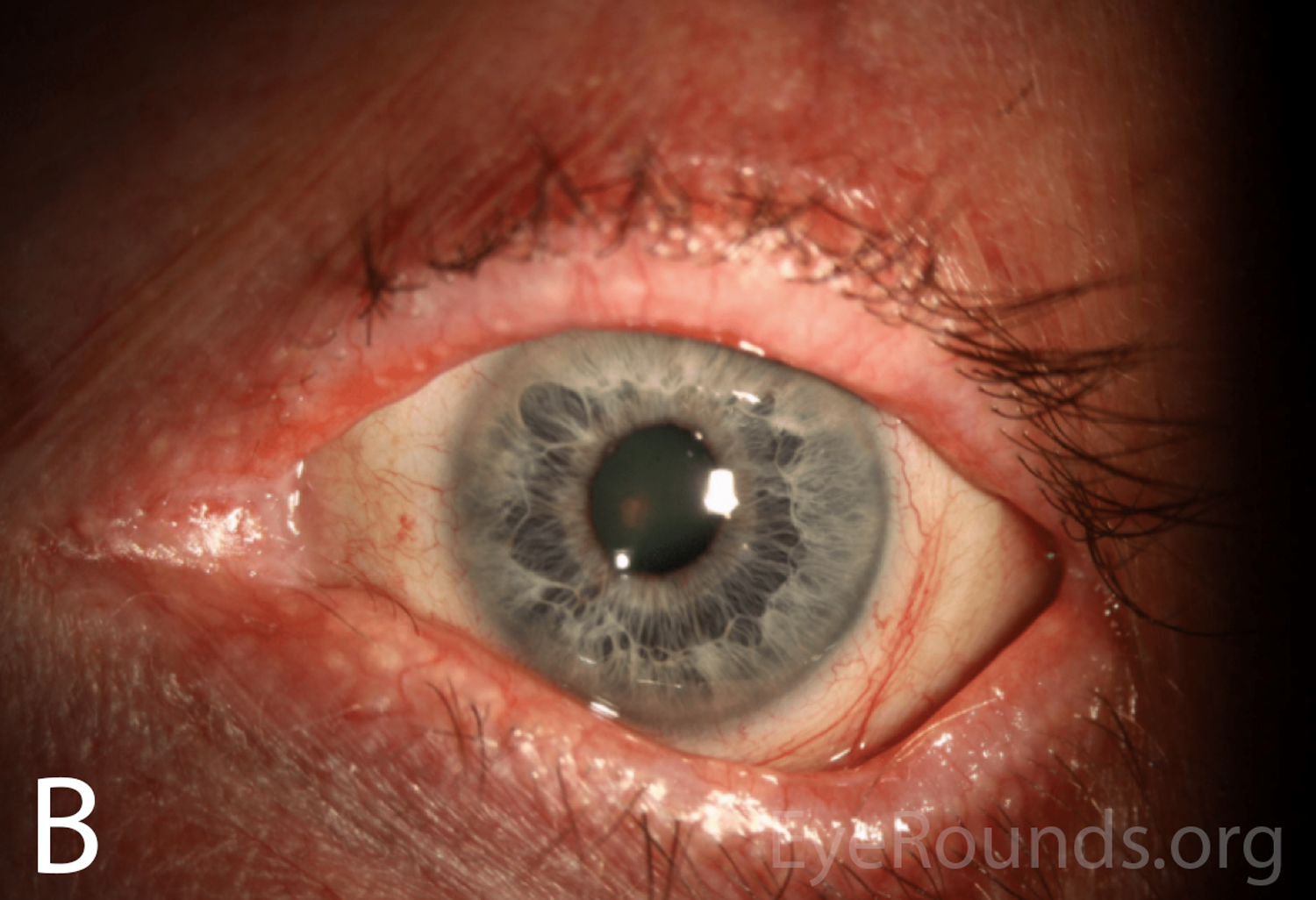
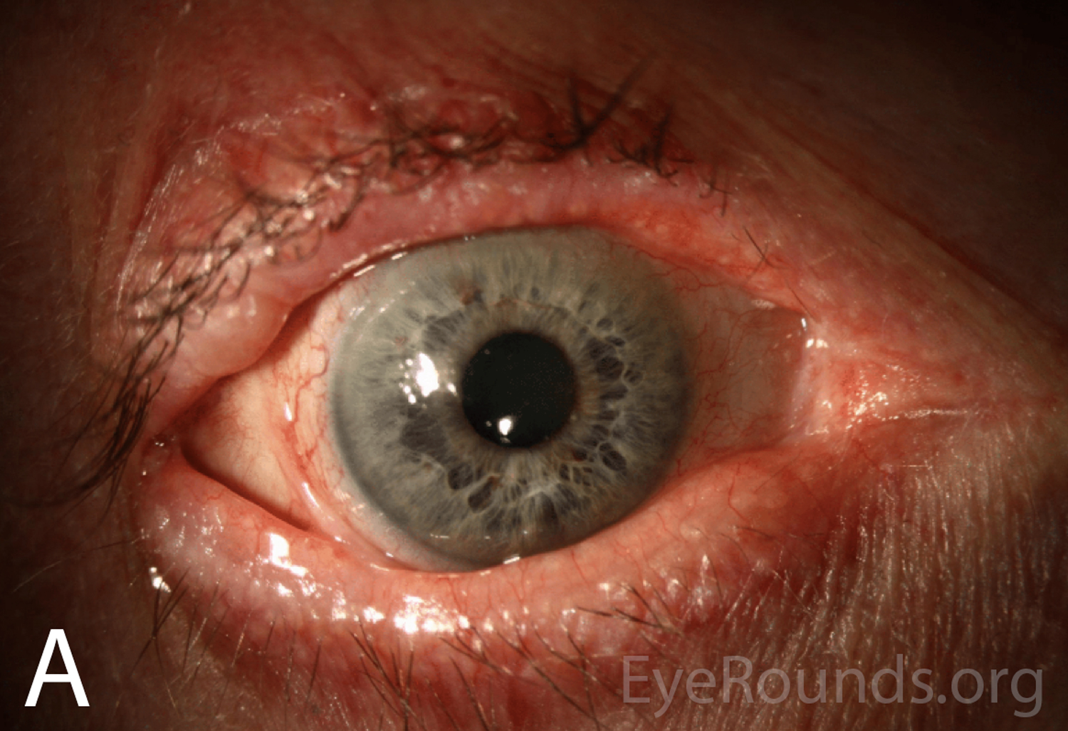
Figure 1A-B. Wide-field slit lamp photographs of the right and left eyes at initial presentation. Note the irregularity and thickening of the eyelid margins, eyelid telangiectasias, punctal fibrosis, MGD, diffuse injection, bulbar conjunctival banding temporally consistent with symblepharon, and keratinization of the plica and caruncle nasally. There is superior corneal pannus in the right eye (A).

Slit lamp photographs of the right and left eyes at initial presentation. In both eyes, note diffuse epithelial haze and scattered punctate epithelial erosions. Slit lamp photographs of the right and left eyes at initial presentation. In both eyes, note diffuse epithelial haze and scattered punctate epithelial erosions.

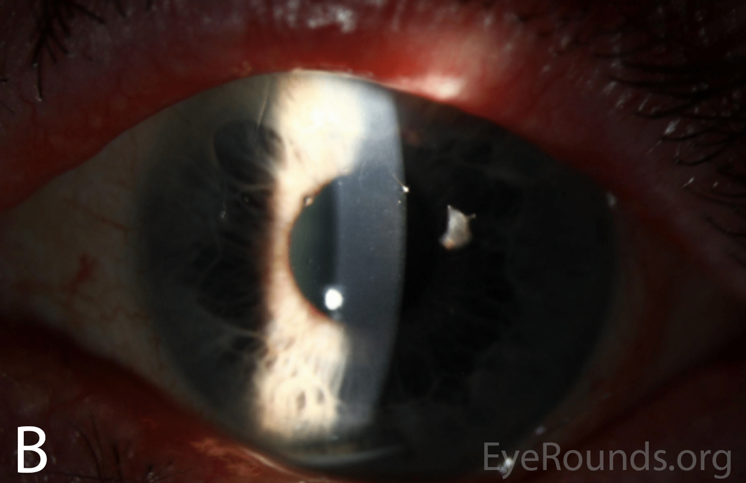
Figure 2A-B. Slit lamp photographs of the right and left eyes at initial presentation. In both eyes, note diffuse epithelial haze and scattered punctate epithelial erosions.

Dilated fundus examination

Normal disc, macula, vessels, and periphery, OU.

Differential Diagnosis

  • Ocular cicatricial pemphigoid
  • Chronic graft vs. host disease
  • Atopic keratoconjunctivitis
  • Sarcoidosis
  • Sjogren's disease related keratoconjunctivitis sicca

CLINICAL COURSE

Prior to his referral to the University of Iowa Hospitals & Clinics (UIHC) Department of Ophthalmology and Visual Sciences, the patient had dry eye, redness, photophobia, and decreased visual acuity that followed a waxing and waning course over a period of eight years. He was repeatedly treated with oral doxycycline, topical steroids, and aggressive lubrication, which seemed to improve his symptoms and vision marginally. He was ultimately referred to UIHC for worsening bilateral symblephara despite topical steroid treatment, which raised concern for ocular cicatricial pemphigoid (OCP).

On initial presentation at UIHC, his exam was thought to be most consistent with chronic GVHD or OCP, based on the presence of bilateral symblephara, buccal mucosal ulcerations, and dental caries. At that time, he was continued on topical prednisolone acetate 1% drops three times daily OU, as well as aggressive lubrication with preservative-free artificial tears every two hours and nightly lubricating gel. He began using autologous serum eye drops (ASEDs) the following month.
Upon follow up, his symptoms and exam were minimally improved on the above regimen.  Conjunctival biopsies were obtained and showed "epithelial changes and scarring most consistent with chronic GVHD." Immunofluorescent studies did not support a diagnosis of ocular cicatricial pemphigoid.

Given the biopsy results, a diagnosis of chronic GVHD was thought most likely. His prednisolone acetate 1% was increased to four times daily and ASEDs were continued every two hours in both eyes. Over the next two months his surface remained stable, with occasional blurred vision and recurrent trichiasis. He was continued on this regimen with regular follow up every three months to monitor disease progression, intraocular pressure, secondary cataract progression, and consideration of adjunctive topical cyclosporine.

DIAGNOSIS

Chronic graft vs. host disease

DISCUSSION

Chronic GVHD is a systemic disease that can occur any time after an allogenic hematopoietic stem cell transplant is performed. The hallmark of chronic GVHD is widespread fibrosis, collagen deposition, and auto-antibody production that commonly affects the skin, lungs, and mucus membranes, which can include the ocular surface and associated secretory glands [1]. This disease entity can be contrasted with acute GVHD, which is an illness that more commonly occurs in the immediate post-transplant period. The diagnostic criteria for chronic GVHD were established in 2005 by a National Institute of Health Consensus Conference and most recently revised in 2014 [2]. 

Epidemiology

Chronic GVHD is a major complication of allogenic hematopoietic stem cell transplantation, with 37% of patients developing the disease within the first year [3] and a cumulative incidence of 64% [4]. Ocular involvement is common, with a large retrospective study finding 33.3% of patients receiving HSCT develop ocular disease although values up to 60-90% have also been reported [5-7]. Notable risk factors for the development of chronic GVHD include prior acute GVHD, peripheral blood stem cell transplants (PBSCT), female donor to male recipient transplant, and EBV-seropositive donor [5]. The disease was once defined as occurring greater than 100 days after transplant, but now it is recognized that chronic GVHD can occur at any time after HSCT [2].

Chronic GVHD can be compared with acute GVHD, a severe inflammatory illness that presents early in the post-transplant period, often while the patient is still hospitalized [8]. Acute GVHD is slightly less common than chronic GVHD, with a reported cumulative incidence of 20% [4]. The acute form of the disease less commonly involves the eye, with a reported incidence of 1.33% in a recent retrospective study [5]. Additionally, the presence of ocular involvement in acute GVHD is a poor prognostic factor, with a mortality rate of approximately 90% [8].

Pathophysiology

Both forms of ocular GVHD are driven by T-cell-mediated inflammatory damage. However, the pathways by which T-cells incite inflammation in the two forms are thought to be different. The exact mechanism of chronic GVHD is poorly understood but the hallmark of the disease is increased IFN-g production [9], which causes a presentation similar to other autoimmune conditions [10]. There is a notable loss of central immune tolerance, resulting in self-reactive T- and B-cells in the periphery [11]. As donor stem cells interact with host T-cells, it is proposed that type 2 T-helper cells produce increased levels of cytokines including TGF-b, although this has only been demonstrated in animal models [12]. This cytokine production results in a reduction in regulatory T-cells and an increase in type 17 T-helper cells, causing widespread T-cell-mediated damage, auto-antibody production, and tissue fibrosis [13, 14]. Inflammatory injury from chronic GVHD commonly affects the skin, lungs, and mucus membranes, but can affect any organ system in the body [15].

Chronic ocular GVHD most commonly presents as keratoconjunctivitis sicca secondary to destruction of the conjunctiva, loss of goblet cells, fibrosis of the lacrimal glands, and meibomian duct fibrosis [16]. These fibrotic changes occur as pro-inflammatory mediators and type 2 T-helper cells result in mononuclear infiltrates, epithelial destruction, and fibrogenesis at these sites [17]. Patients develop severe dry eyes with increased ocular surface inflammation, impaired tear production, and rapid tear evaporation. The impairment of the ocular surface by inflammation can further lead to complications such as limbal stem cell deficiency, corneal scarring, symblepharon, entropion, and trichiasis [8, 18].

Disease mechanisms are better characterized in acute GVHD, a disease in which recipient antigens are attacked directly by donor T-lymphocytes [19]. The organs most commonly affected are skin, causing desquamation and bulla formation; liver, causing cholestatic disease and hyperbilirubinemia; and bowel epithelium, causing severe diarrhea [2]. Given the skin manifestations, patients may present similarly to those with toxic epidermal necrolysis [20].

Signs/Symptoms

The defining feature of chronic ocular GVHD is one of severe dry eye with conjunctival inflammation; symptoms occur on a spectrum. The most commonly reported symptoms are bilateral eye irritation, pain, burning sensation, photophobia, redness, blurred vision, and/or foreign body sensation [16].

Common exam findings include MGD, rapid tear break up time, decreased tear production, punctate epitheliopathy, conjunctival hyperemia, and corneal haze. As the disease progresses and surface inflammation increases, there can be development of corneal filaments, corneal scarring, cicatricial entropion, trichiasis, and/or symblepharon [21-23]. The exam may also reveal periadnexal dermatitis and vitiligo [23]. Late complications can include cataract, uveitis, retinal microvasculopathy with cotton-wool spots, vitreous hemorrhage, and/or optic disc edema [24].

Although the eyes are not widely considered a target organ for acute GVHD, when it does occur the hallmark of acute ocular GVHD is a pseudomembranous conjunctivitis with epithelial sloughing.  Though, the most common ocular symptoms are also those of dry eye. These patients are often critically ill with other systemic complications [8].

Testing

Diagnosis of ocular GVHD is made clinically based upon degree of clinical suspicion in patients with keratoconjunctivitis and a history of allogenic HSCT. The Center for International Blood and Marrow Transplant Research published a review on ocular GVHD which included recommended evaluation measures for dry eye severity [25]:

  • Schirmer testing for aqueous deficiency – For Schirmer I testing without anesthesia, tear production <10 mm in 5 minutes is reported to have a sensitivity and specificity of 80%, although this is not specific to GVHD and does not account for tear evaporation as a cause of dry eye [21, 22].
  • Ocular surface disease index – A questionnaire on dry eye related symptoms can be administered by hematologists to screen for dry eye with a sensitivity of 44% and specificity of 98% [21, 26].
  • Fluorescein corneal staining – Evaluating for corneal epithelial pathology in the setting of GVHD has been shown to have a sensitivity of 91% and specificity of 54% [21, 22].
  • Tear film breakup time (TBUT) – A rapid TBUT of less than 5 seconds is reported to have a sensitivity of 80% and specificity of 67% [21, 22].

Dry eye symptoms can be graded from 0 - 3 using the NIH Organ Scoring system, which is a standardized and validated tool that grades chronic GVHD severity by organ system [2, 27].

  • GVHD Score 0 – No dry eye symptoms (DES).
  • GVHD Score 1 – DES not affecting activities of daily living (ADL) and requiring eye drops less than 3 times per day.
  • GVHD Score 2 – DES that partially affect ADL and require eye drops more than 3 times per day or punctal plugs; without vision impairment.
  • GVHD Score 3 – DES that significantly affect ADL, render the patient unable to work, or cause a loss of vision.

Although not required for the diagnosis of ocular GVHD, a conjunctival biopsy can show findings that are supportive of chronic GVHD. These changes include epithelial keratinization, a decrease in goblet cells, vacuolar alteration of basal cells, epithelial atrophy, and scarring from chronic disease [28]. A biopsy can also be useful when the diagnosis is not clear, or when there is concern for other pathologies such as OCP.  

Management

The National Institutes of Health (NIH) released an updated consensus statement on the management of chronic GVHD in 2015 with systems-focused recommendations of disease management, of which the complete guidelines are beyond the scope of this review [15]. They identified four supportive care goals for chronic ocular GVHD: lubrication, control of tear evaporation, control of tear drainage, and decreasing surface inflammation [15].

Increased lubrication can be achieved through the judicious use of preservative-free artificial tears and nightly lubricating gels or ointments [29]. Although the NIH report lists oral muscarinic agonists such as cevimeline  and pilocarpine  as potential treatments due to their use in other dry eye syndromes [30, 31], there is little evidence of their efficacy in GVHD. Tear evaporation can be improved in the setting of MGD with warm compresses [32] and oral doxycycline [33], as well as through the use of moisture chamber goggles or scleral lenses [15, 34]. Tear drainage can be reduced through the use of temporary or permanent punctal occlusion [35].

There are a variety of ways to decrease surface inflammation, with limited evidence available to support the efficacy of specific therapies. The current first-line treatment is topical steroids, which non-specifically decrease ocular surface inflammation [36]. One retrospective study found that topical prednisolone acetate 1% drops produced a complete resolution of symptoms with a mean therapy duration of seven weeks [36]. Concerns with topical steroid use include the potential for development of cataracts, steroid-induced ocular hypertension, and increased risk of infectious keratitis [37]. Topical cyclosporine-A 0.05% has also been shown to decrease inflammation with significantly fewer side effects than steroids by inhibiting T-cell activation and downregulating inflammatory cytokine levels in the conjunctiva, although it appears to be less efficacious [38]. The NIH report recommended higher doses, such as cyclosporine 0.1%, to decrease surface inflammation [15]. There is also strong evidence that ASEDs used every two hours can produce a notable decrease in surface inflammation by a number of mechanisms including supplying platelet derived growth factor [39] resulting in improved visual acuity, improvement in symptoms, and decreased need for artificial tears [40]. If topical or local therapies are no longer effective, the patient's hematologist/oncologist should be consulted for consideration of systemic immunosuppressive therapy [41].

Acute GVHD is less likely to be encountered by the ophthalmologist, as it often occurs in the immediate post-transplant period in a severely ill patient [8]. The treatment depends on the specific organ systems involved, but the mainstay of treatment of severe systemic disease including in cases of ocular involvement is T-lymphocyte suppression. Most commonly, this will be performed with methylprednisolone and adjustment of the patient's current immunosuppressive therapy [42].

EPIDEMIOLOGY
  • Occurs any time after allogenic HSCT
  • Incidence of up to 64%
  • Ocular involvement in up to 90% of cases
  • Increased risk with acute GVHD, PBSCT, female to male transplant, and EBV-seropositive donors
SIGNS
  • Punctate epitheliopathy
  • Decreased Schirmer I testing
  • Conjunctival hyperemia
  • Meibomian gland dysfunction
  • Rapid TBUT
  • Corneal haze
  • Corneal filaments
  • Corneal scarring
  • Cicatricial entropion
  • Trichiasis
  • Symblepharon
SYMPTOMS
  • Eye redness
  • Eye irritation
  • Blurry vision
  • Foreign body sensation
  • Eye pain or burning
  • Photophobia
MANAGEMENT
  • Preservative-free artificial tears
  • Nightly lubricating ointments
  • Warm compresses
  • Punctal occlusion
  • Oral doxycycline
  • ASEDs
  • Scleral lenses
  • Topical steroids
  • Topical cyclosporine
  • Systemic immunosuppression
References
  1. Ferrara J, Levine J, Reddy P, Holler E. Graft-versus-host disease. Lancet 2009;373(9674):1550-1561. 
  2. Jagasia M, Greinix H, Arora M, Williams K, Wolff D, Cowen E, Palmer J, Weisdorf D. National Institutes of Health Consensus Development Project on Criteria for Clinical Trials in Chronic Graft-versus-Host Disease: I. The 2014 Diagnosis and Staging Working Group report. Biol Blood Marrow Transplant 2015;21(3):389-401. 
  3. Arai S, Arora M, Wang T, Spellman SR, He W, Couriel DR, Urbano-Ispizua A, Cutler CS, Bacigalupo AA, Battiwalla M, Flowers ME, Juckett MB, Lee SJ, Loren AW, Klumpp TR, Prockup SE, Ringden OT, Savani BN, Socie G, Schultz KR, Spitzer T, Teshima T, Bredeson CN, Jacobsohn DA, Hayashi RJ, Drobyski WR, Frangoul HA, Akpek G, Ho VT, Lewis VA, Gale RP, Koreth J, Chao NJ, Aljurf MD, Cooper BW, Laughlin MJ, Hsu JW, Hematti P, Verdonck LF, Solh MM, Norkin M, Reddy V, Martino R, Gadalla S, Goldberg JD, McCarthy PL, Perez-Simon JA, Khera N, Lewis ID, Atsuta Y, Olsson RF, Saber W, Waller EK, Blaise D, Pidala JA, Martin PJ, Satwani P, Bornhauser M, Inamoto Y, Weisdorf DJ, Horowitz MM, Pavletic SZ, Graft-vs-Host Disease Working Committee of the C. Increasing incidence of chronic graft-versus-host disease in allogeneic transplantation: a report from the Center for International Blood and Marrow Transplant Research. Biol Blood Marrow Transplant 2015;21(2):266-274.  https://PubMed.gov/25445023. DOI: 10.1016/j.bbmt.2014.10.021
  4. Khouri IF, Saliba RM, Giralt SA, Lee MS, Okoroji GJ, Hagemeister FB, Korbling M, Younes A, Ippoliti C, Gajewski JL, McLaughlin P, Anderlini P, Donato ML, Cabanillas FF, Champlin RE. Nonablative allogeneic hematopoietic transplantation as adoptive immunotherapy for indolent lymphoma: low incidence of toxicity, acute graft-versus-host disease, and treatment-related mortality. Blood 2001;98(13):3595-3599.  https://PubMed.gov/11739162. DOI: 10.1182/blood.v98.13.3595
  5. Na K, Yoo Y, Mok J, Lee J, Joo C. Incidence and risk factors for ocular GVHD after allogeneic hematopoietic stem cell transplantation. Bone Marrow Transplant 2015;50(11):1459-1464. 
  6. Anderson NG, Regillo C. Ocular manifestations of graft versus host disease. Curr Opin Ophthalmol 2004;15(6):503-507.  https://PubMed.gov/15523196
  7. Hessen M, Akpek EK. Ocular graft-versus-host disease. Curr Opin Allergy Clin Immunol 2012;12(5):540-547.  https://PubMed.gov/22892710. DOI: 10.1097/ACI.0b013e328357b4b9
  8. Jabs DA, Wingard J. The Eye in Bone Marrow Transplantation: Conjuctival Graft-vs-Host Disease. Arch Ophthalmology 1989;107:1343-1348. 
  9. Cavet J, Dickinson AM, Norden J, Taylor PR, Jackson GH, Middleton PG. Interferon-gamma and interleukin-6 gene polymorphisms associate with graft-versus-host disease in HLA-matched sibling bone marrow transplantation. Blood 2001;98(5):1594-1600.  https://PubMed.gov/11520812. DOI: 10.1182/blood.v98.5.1594
  10. Gilman A, Serody J. Diagnosis and treatment of chronic graft-versus-host disease. Semin Hematol 2006;43(1):70-80. 
  11. Min C-K. The pathophysiology of chronic graft-versus-host disease: the unveiling of an enigma. Korean J Hematol 2011;46(2):80-87. 
  12. McCormick L, Zhang Y, Tootell E, Gilliam A. Anti-TGF-beta treatment prevents skin and lung fibrosis in murine sclerodermatous graft-versus-host disease: a model for human scleroderma. J Immunol 1999;163(10):5693-5699. 
  13. Choi S, Levine J, Ferrara J. Pathogenesis and Management of Graft-versus-Host Disease. Immunol Allergy Clin North Am 2010;30(1):75-101. 
  14. Dander E, Balduzzi A, Zappa G, Lucchini G, Perseghin P, Andre V, Todisco E, Rahal D, Migliavacca M, Longoni D, Solinas G, Villa A, Berti E, Mina PD, Parma M, Allavena P, Biagi E, Rovelli A, Biondi A, D'Amico G. Interleukin-17-producing T-helper cells as new potential player mediating graft-versus-host disease in patients undergoing allogeneic stem-cell transplantation. Transplantation 2009;88(11):1261-1272.  https://PubMed.gov/19996925. DOI: 10.1097/TP.0b013e3181bc267e
  15. Carpenter PA, Kitko CL. National Institutes of Health Consensus Development Project on Criteria for Clinical Trials in Chronic Graft-versus-Host Disease: V. The 2014 Ancillary Therapy and Supportive Care Working Group Report. Biol Blood Marrow Transplant 2015;21:1167-1187. 
  16. Jacobs R, Tran U, Chen H, Kassim A, Engelhardt B, Greer J, Goodman S, Clifton C, Lucid C, Vaughan L, Savani B, Jagasia M. Prevalence and risk factors associated with development of ocular GVHD defined by NIH consensus criteria. Bone Marrow Transplant 2012;47(11):1470-1473. 
  17. Zhang C, Todorev I, Zhang Z, Liu Y, Kandeel F, Forman S, Strober S, Zeng D. Donor CD4+ T and B cells in transplants induce chronic graft-versus-host disease with autoimmune manifestations. Blood 2005;107(7):2993-3001. 
  18. Marzano A, Facchetti M, Berti E, Caputo R. Chronic graft-vs.-host disease with severe cicatrizing conjunctivitis mimicking cicatricial pemphigoid. Br J Ophthalmol 2000;143(1):209-210. 
  19. Ferrara J, Reddy P. Pathophysiology of graft-versus-host disease. Semin Hematol 2006;43(1):3-10.  
  20. Naik H, Lockwood S, Saavedra A. A pilot study comparing histological and immunophenotypic patterns in stage 4 skin graft vs host disease from toxic epidermal necrolysis. J Cutan Pathol 2017;44(10):857-860. 
  21. Pathak M, Diep P, Lai X, Brinch L, Ruud E, Drolsum L. Ocular findings and ocular graft-versus-host disease after allogeneic stem cell transplantation without total body irradiation. Bone Marrow Transplant 2018;53(7):863-872. 
  22. Curtis L, Datiles M, Steinberg S, Mitchell S, Bishop R, Cowen E, Mays J, McCarty J, Kuzmina Z, Pirsl F. Predictive models for ocular chronic graft-versus-host disease diagnosis and disease activity in transplant clinical practice. Haematologica 2015;100(9):1228-1236. 
  23. Hessen M, Akpek E. Ocular graft-versus-host disease. Curr Opin Allergy Clin Immunol 2012;12(5):540-547. 
  24. Coskuncan N, Jabs D, Dunn J, Haller J, Green W, Vogelsang G, Santos G. The eye in bone marrow transplantation. VI. Retinal complications. Arch Ophthalmology 1994;112(3):372-379. 
  25. Inamoto Y, Valdes-Sanz N, Ogawa Y, Alves M, Berchicci L, Galvin J, Greinix H, Hale G. Ocular graft-versus-host disease after hematopoietic cell transplantation: Expert review from the Late Effects and Quality of Life Working Committee of the CIBMTR and Transplant Complications Working Party of the EBMT. Biol Blood Marrow Transplant 2019;25(2):e46-e54. 
  26. Inamoto Y, Chai X, Kurland B, Cutler C, Flowers M, Palmer J, Carpenter P, Heffernan M. Validation of measurement scales in ocular graft-versus-host disease. Ophthalmology 2012;119(3):487-493. 
  27. Filipovich A, Weisdorf D, Pavletic S, Socie G, Wingard J, Lee S, Martin P, Chien J. National Institutes of Health Consensus Development Project on Criteria for Clinical Trials in Chronic Graft-versus-Host Disease: I. Diagnosis and Staging Working Group Report. Biol Blood Marrow Transplant 2005;11(12):945-956. 
  28. Auw-Haedrich C, Potsch C. Histological and immunohistochemical characterisation of conjunctival graft vs host disease following haematopoietic stem cell transplantation. Graefe's Arch Clin Exp Ophthalmol 2007;245:1001-1007. 
  29. Behrens A, Doyle J, Stern L, Chuck R, McDonnell P, Azar D, Dua H, Hom M. Dysfunctional tear syndrome: a Delphi approach to treatment recommendations. Cornea 2006;25(8):900-907. 
  30. Papas A, Sherrer Y, Charney M, Golden H, Medsger T, Walsh B, Trivedi M, Goldlust B. Successful treatment of dry mouth and dry eye symptoms in Sjogren's syndrome patients with oral pilocarpine: a randomized, placebo-controlled, dose-adjustment study. J Clin Rheumatol 2004;10(4):169-177. 
  31. Ono M, Takamura E, Shinozaki K, Tsumura T, Hamano T, Yagi Y, Tsubota K. Therapeutic effect of cevimeline on dry eye in patients with Sjogren's syndrome: a randomized, double-blind clinical study. Am J Ophthalmol 2004;138(1):6-17. 
  32. Olson M, Korb D, Greiner J. Increase in tear film lipid layer thickness following treatment with warm compresses in patients with meibomian gland dysfunction. Eye Contact Lens 2003;29(2):96-99. 
  33. Frucht-Pery J, Sagi E, Hemo I, Ever-Hadani P. Efficacy of doxycycline and tetracycline in ocular rosacea. Am J Ophthalmol 1993;116(1):88-92. 
  34. Jacobs D, Rosenthal P. Boston scleral lens prosthetic device for treatment of severe dry eye in chronic graft-versus-host disease. Cornea 2007;26(10):1195-1199. 
  35. Sabti S, Halter J, Braun Frankl B, Goldblum D. Punctal occlusion is safe and efficient for the treatment of keratoconjunctivitis sicca in patients with ocular GvHD. Bone Marrow Transplant 2012;47(7):981-984. 
  36. Robinson M, Lee S. Topical corticosteroid therapy for cicatricial conjunctivitis associated with chronic graft-versus-host disease. Bone Marrow Transplantation 2004;33:1031-1035. 
  37. McGhee C, Dean S, Danesh-Meyer H. Locally administered ocular corticosteroids: benefits and risks. Drug Saf 2002;25(1):33-55. 
  38. Baptista Malta J, Soong H, Shtein RM, Musch DC, Rhoades W, Sugar A, Mian S. Treatment of Ocular Graft-Versus-Host Disease With Topical Cyclosporine 0.05%. Cornea 2010;29:1392-1396. 
  39. Pezzotta S, Del Fante C, Scudeller L, Cervio M, Antoniazzi E, Perotti C. Autologous platelet lysate for treatment of refractory ocular GVHD. Bone Marrow Transplant 2012;47(12):1558-1563. 
  40. Tahmaz V, Gehlsen U. Treatment of severe chronic ocular graft-versus-host disease using 100% autologous serum eye drops from a sealed manufacturing system: a retrospective cohort study. Br J Ophthalmol 2017;101:322-326. 
  41. Nassiri N, Eslani M. Ocular Graft Versus Host Disease Following Allogeneic Stem Cell Transplantation: A Review of Current Knowledge and Recommendations. J Ophthalmic Vis Res 2013;8(4):351-358. 
  42. Hings I, Severson R, Filipovich A, Balazar B, Kersey J, Ramsay N, McGalve P, Weisdorf D. Treatment of moderate and severe acute GVHD after allogeneic bone marrow transplantation. Transplantation 1994;58(4):437-442. 
Suggested citation format:

Goldstein AS, Wilson CW, Greiner MA. Chronic Ocular Graft Versus Host Disease. EyeRounds.org. Posted August 8, 2019; Available from https://EyeRounds.org/cases/287-GVHD.htm


last updated: 08/08/2019

Image Permissions:
Creative Commons

Ophthalmic Atlas Images by EyeRounds.org, The University of Iowa are licensed under a Creative Commons Attribution-NonCommercial-NoDerivs 3.0 Unported License.