The COMS Grading Scheme: Graded Features
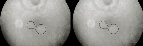
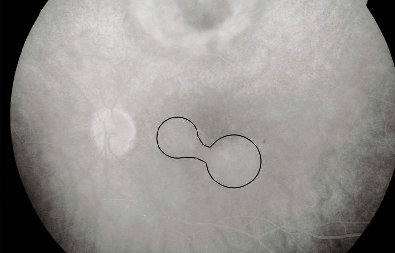
Macular Angiographic Leakage
(Macular edema)
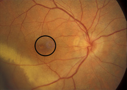
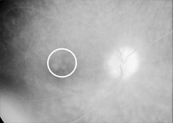
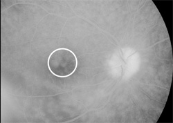
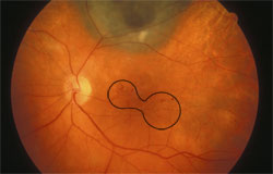
The wide-field photographic techniques used in the COMS rarely provide a stereoscopic image to determine retinal edema and thickening. Surrogate markers are used. Macular edema often causes mild opacification of the involved retina which appears slightly milky in comparison to the surrounding retina. On angiography the retinal capillary bed is often abnormal with microaneurysms, areas of capillary non-perfusion, or diffusely dilated capillaries. In the late frames of the angiogram, hyperfluorescence is seen in the macular region, often taking on a petalloid pattern, or a pattern of finely clustered circles.
Severity
back to COMS index