The COMS Grading Scheme: Graded Features
Laser scars
(focal or pan-retinal photocoagulation)
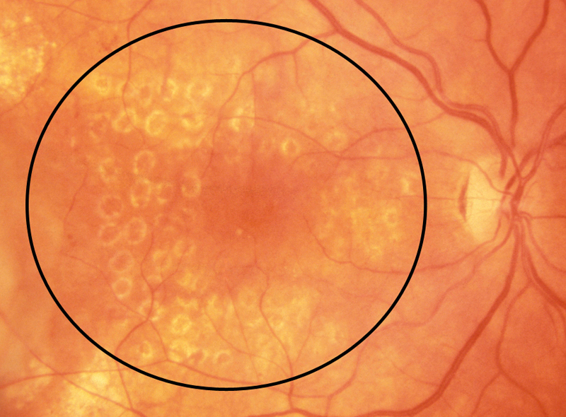
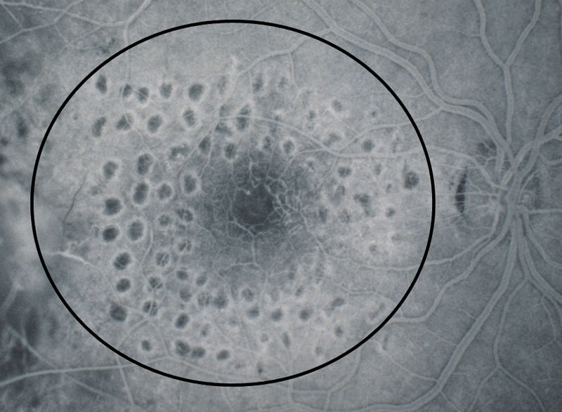
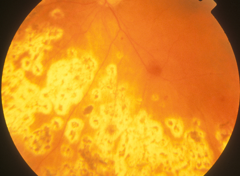
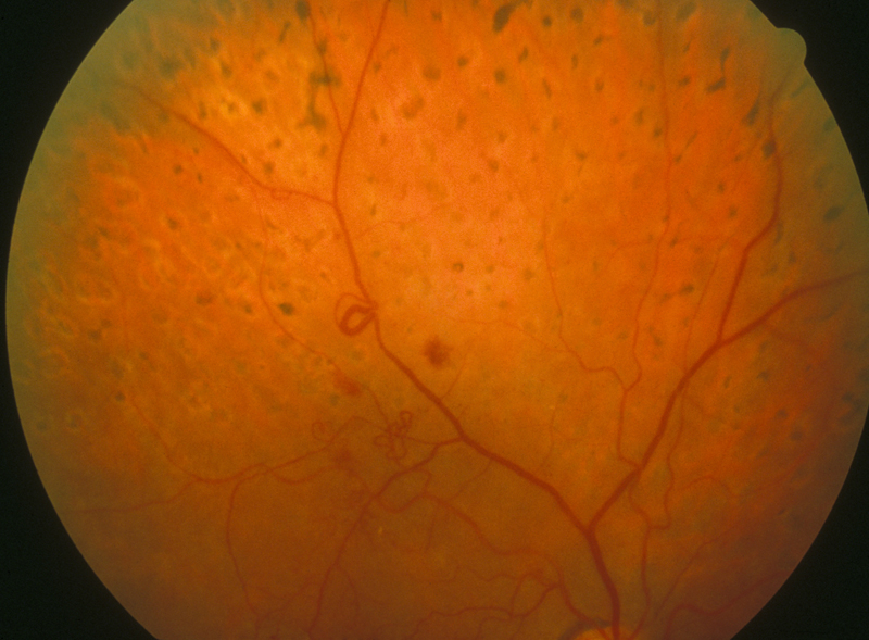
Laser scars characteristically appear as 50 to 200 micron diameter lesions in the macula or 300-600 micron lesions in the periphery. They are round or oval, yellowish-white with variable black pigment centrally. Focal laser is defined as laser placed within the macula. Scatter laser is defined as more extensive laser, the majority of which is placed outside the macula. If the laser scars were in the photograph but not in the macular or peripapillary fields, they were graded as present.
Severity
| click on any image for higher resolution image click on your browser's "back" button to return to this page |
||
| None: not present | ||
Focal: laser placed within the macula (standard 1 with angiogram)
|
||
Scatter: more extensive laser, the majority of which was outside the macula (standards 2 and 3)
|
back to COMS index