The COMS Grading Scheme: Graded Features
Retinal Hemorrhage
All punctuate, blot, or linear hemorrhages are included. Hemorrhages that are clearly subretinal or preretinal are excluded. Retinal hemorrhages show blocked fluorescence on angiogram. If an angiogram is not available, red dots on color photographs are called retinal hemorrhages unless the grader feels they represent microaneurysms.
Severity
| None: not present |
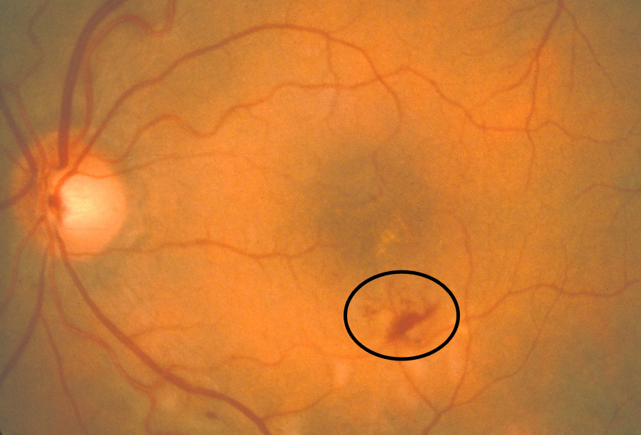
Mild: total area less than 1/5 disc area (standard photograph 1)
|
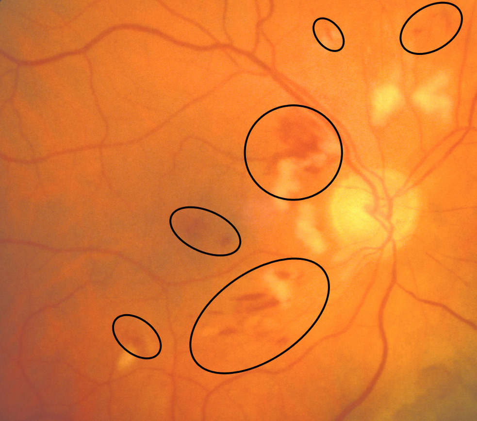
Moderate: total area more than 1/5 disc area, but less than 1 disc area (standard photograph 2)
|
| Severe: total area more than 1 disc area |
back to COMS index