The COMS Grading Scheme: Graded Features
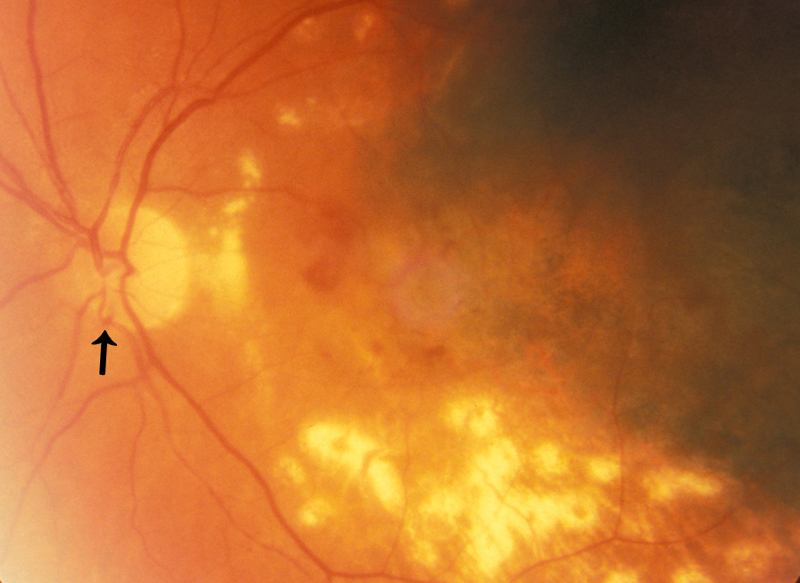
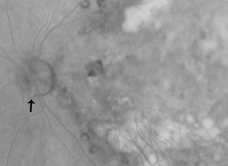
Optic disc hemorrhage
Optic disc hemorrhage is often linear, following the pattern of the nerve fiber layer. It is located anterior or within the superficial tissue of the optic nerve head. If a hemorrhage starts over the optic nerve head but extends into the peripapillary retina, it is still considered an optic nerve hemorrhage. The grading is based on both the area of the optic disc covered by the hemorrhage and the number of hemorrhages.
Severity
| click on any image for higher resolution image click on your browser's "back" button to return to this page |
|||||
| None: not present | |||||
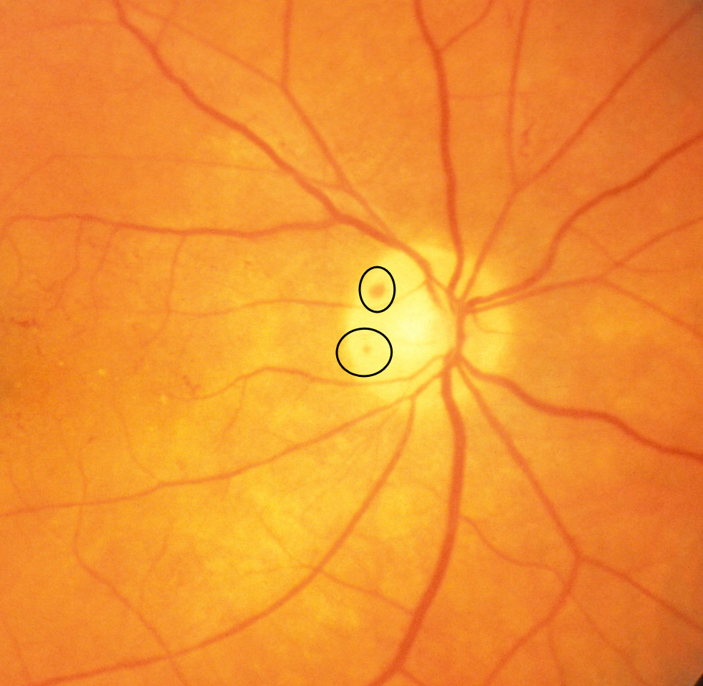
Mild: 1 or 2 hemorrhages of less than 1/8 disc area combined (standard 1)
|
|||||
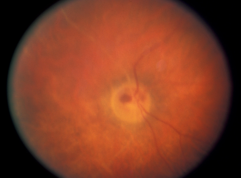
Moderate: 1 or 2 hemorrhages of 1/8 disc area or more but less than ¼ disc area, or 3 to 4 hemorrhages of less than ¼ disc area combined (standard 2 and 3)
|
|||||
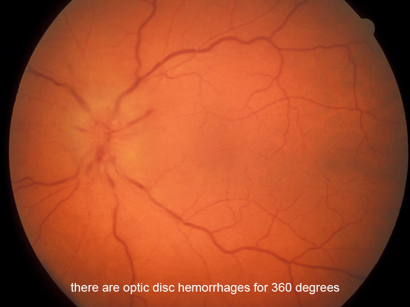
Severe: 5 or more hemorrhages or total area ¼ disc area or more (standard 4)
|
back to COMS index