Images from the Laboratory of Dr. Rob Mullins
 |
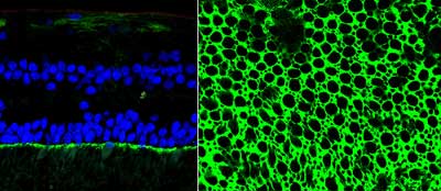
Two views of ICAM-1 in the external limiting membrane and/or Muller cell microvilli. The left image is an epifluorescence micrograph of a sectioned human retina labeled with an antibody directed against ICAM-1 (green) and depicted with a nuclear counterstain (blue). The right image is a confocal micrograph in which the retina is rotated 90 degrees compared with the image on the left, such that the viewer is looking down on the external limiting membrane. Note the honeycomb-like distribution of ICAM-1. Cone and rod photoreceptor cells traverse this layer through its large and small openings. We recently described regional variation in ICAM-1 expression in the choriocapillaris and external limiting membrane of human eyes (Mullins et al., Molecular Vision 12:224-35). |
click on image for higher resolution image |
 |
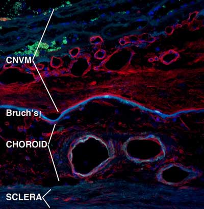
Endothelial cells at different stages of differentiation alter their cell surface carbohydrates (sugars that are associated with glycoproteins or glycolipids). These carbohydrate molecule changes can be detected with lectins—plant, animal or fungal proteins that physically interact with different, specific sugar groups. This image shows lectin labeling (red) of blood vessels and other structures in a human eye with a choroidal neovascular membrane (CNVM). Choroidal neovascularization is a frequent end-stage of age-related macular degeneration in which blood vessels from the choroid grow through Bruch’s membrane. Blood vessels appear as circles or ovals. Note the robust labeling of the vessels within the CNVM, as compared with the minor labeling of “normal” vessels in the choroid. Cell nuclei are counterstained blue. We recently described patterns of carbohydrate molecules in eyes with macular degeneration (Mullins et al., 2005, Molecular Vision 11:509-17). |
click on image for higher resolution image |

