Gonioscopy is one of the more challenging eye examination techniques for ophthalmology residents to learn. Unfortunately, in clinical practice gonioscopy is not performed as often as it should be. In a study of 193 primary open angle glaucoma charts from a community-based sample in the United States, only 51.3% of patients had gonioscopy performed at initial presentation (Hertzog LH, Albrecht KG, LaBree L, Lee PP. Ophthalmology 1996;103:1009-13). Gonioscopy is a critical part of the eye examination. There is much to be seen by gonioscopy – not simply whether the iridocorneal angle is open or closed
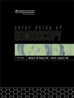
 This website is meant to share gonioscopy techniques and images. In 1994 I wrote a textbook on gonioscopy entitled Color Atlas of Gonioscopy that was published by Wolfe (an imprint of Mosby). In 2001 the American Academy of Ophthalmology reprinted this textbook. A second edition was published with the co-authorship of Reid A. Longmuir, M.D. in 2008. The Atlas is available through the American Academy of Ophthalmology at http://www.aao.org. In the spirit of full disclosure I should point out that, because this book is now published by the American Academy of Ophthalmology, I receive no royalties for any copies sold.
While drawings, paintings and photographs of the iridocorneal angle are helpful for the student of gonioscopy, I have found that videography demonstrates the techniques and findings much more readily than any other medium. This site is therefore designed to teach how to perform gonioscopy and also to show the iridocorneal angle in health and in disease. There are occasional slit lamp videos as well if they provide illustrations of the findings in glaucoma-related diseases.
Too often only the most extreme examples of disease are used as examples. For the novice, however, it is the subtle examples of disease that are the most puzzling. Being unconstrained by space, my hope is that this site will show the dramatic polycoria seen in the iridocorneal endothelial syndrome but also the early form of the disease in which one may only see a single synechia.
Please recognize that the opinions regarding preferred lenses and grading systems expressed are my own. Residents and fellows should be aware that my opinions might be different than your mentors and, since your mentors will be evaluating you, you should listen to them.
I am grateful to several people for their help with this project. My partners Young H. Kwon, MD, PhD, John H. Fingert, MD, PhD, Nathanial C. Sears, MD, Erin A. Boese, MD and Andrew Pouw, MD have contributed some of these videos and have made the Glaucoma Service a place that celebrates teaching and learning.
Lastly, I am especially grateful to Randall Verdick and Jessica Bramow. Randy Verdick taught me how to capture and edit video. Jessica Bramow designed and built this webpage. She has demonstrated tremendous creativity and skill. It has been fun working on this project mostly because these two have had as much enthusiasm for it as I have.
I hope that you find it useful.
Wallace L.M. Alward, MD
Professor Emeritus
Department of Ophthalmology and Visual Sciences
University of Iowa Carver College of Medicine